5A1A

2.2 A resolution cryo-EM structure of beta-galactosidase in complex with a cell-permeant inhibitor


Experimental Data Snapshot

  • Method: ELECTRON MICROSCOPY
  • Resolution: 2.20 Å
  • Aggregation State: PARTICLE 
  • Reconstruction Method: SINGLE PARTICLE 

wwPDB Validation   3D Report Full Report


This is version 1.4 of the entry. See complete history


Literature

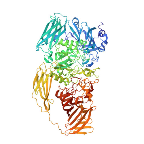
2.2 A Resolution Cryo-Em Structure of Beta-Galactosidase in Complex with a Cell-Permeant Inhibitor

Bartesaghi, A.Merk, A.Banerjee, S.Matthies, D.Wu, X.Milne, J.Subramaniam, S.

(2015) Science 348: 1147

  • DOI: https://doi.org/10.1126/science.aab1576
  • Primary Citation of Related Structures:  
    5A1A

  • PubMed Abstract: 

    Cryo-electron microscopy (cryo-EM) is rapidly emerging as a powerful tool for protein structure determination at high resolution. Here we report the structure of a complex between Escherichia coli β-galactosidase and the cell-permeant inhibitor phenylethyl β-D-thiogalactopyranoside (PETG), determined by cryo-EM at an average resolution of ~2.2 angstroms (Å). Besides the PETG ligand, we identified densities in the map for ~800 water molecules and for magnesium and sodium ions. Although it is likely that continued advances in detector technology may further enhance resolution, our findings demonstrate that preparation of specimens of adequate quality and intrinsic protein flexibility, rather than imaging or image-processing technologies, now represent the major bottlenecks to routinely achieving resolutions close to 2 Å using single-particle cryo-EM.


  • Organizational Affiliation

    Laboratory of Cell Biology, Center for Cancer Research, National Cancer Institute, National Institutes of Health, Bethesda, MD 20892, USA.


Macromolecules
Find similar proteins by:  (by identity cutoff)  |  3D Structure
Entity ID: 1
MoleculeChains Sequence LengthOrganismDetailsImage
BETA-GALACTOSIDASE
A, B, C, D
1,022Escherichia coli K-12Mutation(s): 0 
EC: 3.2.1.23
UniProt
Find proteins for P00722 (Escherichia coli (strain K12))
Explore P00722 
Go to UniProtKB:  P00722
Entity Groups  
Sequence Clusters30% Identity50% Identity70% Identity90% Identity95% Identity100% Identity
UniProt GroupP00722
Sequence Annotations
Expand
  • Reference Sequence
Small Molecules
Ligands 3 Unique
IDChains Name / Formula / InChI Key2D Diagram3D Interactions
PTQ
Query on PTQ

Download Ideal Coordinates CCD File 
E [auth A],
J [auth B],
O [auth C],
T [auth D]
2-phenylethyl 1-thio-beta-D-galactopyranoside
C14 H20 O5 S
ZNAMMSOYKPMPGC-HTOAHKCRSA-N
MG
Query on MG

Download Ideal Coordinates CCD File 
F [auth A]
G [auth A]
K [auth B]
L [auth B]
P [auth C]
F [auth A],
G [auth A],
K [auth B],
L [auth B],
P [auth C],
Q [auth C],
U [auth D],
V [auth D]
MAGNESIUM ION
Mg
JLVVSXFLKOJNIY-UHFFFAOYSA-N
NA
Query on NA

Download Ideal Coordinates CCD File 
H [auth A]
I [auth A]
M [auth B]
N [auth B]
R [auth C]
H [auth A],
I [auth A],
M [auth B],
N [auth B],
R [auth C],
S [auth C],
W [auth D],
X [auth D]
SODIUM ION
Na
FKNQFGJONOIPTF-UHFFFAOYSA-N
Experimental Data & Validation

Experimental Data

  • Method: ELECTRON MICROSCOPY
  • Resolution: 2.20 Å
  • Aggregation State: PARTICLE 
  • Reconstruction Method: SINGLE PARTICLE 
EM Software:
TaskSoftware PackageVersion
RECONSTRUCTIONFREALIGN

Structure Validation

View Full Validation Report


Entry History 

Deposition Data

Revision History  (Full details and data files)

  • Version 1.0: 2015-05-06
    Type: Initial release
  • Version 1.1: 2015-05-13
    Changes: Database references
  • Version 1.2: 2015-05-20
    Changes: Database references
  • Version 1.3: 2015-06-24
    Changes: Database references
  • Version 1.4: 2018-10-03
    Changes: Data collection