


Crystal structure of Escherichia coli ElaA
Shi, L.L., Gao, Z.Q., Zhang, H., Dong, Y.H.To be published.
Experimental Data Snapshot
wwPDB Validation 3D Report Full Report

Entity ID: 1 | |||||
|---|---|---|---|---|---|
| Molecule | Chains | Sequence Length | Organism | Details | Image |
| Protein ElaA | 153 | Escherichia coli K-12 | Mutation(s): 0 Gene Names: elaA, yfbC, b2267, JW2262 | 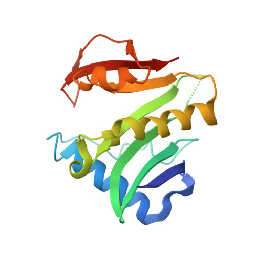 | |
UniProt | |||||
Find proteins for P0AEH3 (Escherichia coli (strain K12)) Explore P0AEH3 Go to UniProtKB: P0AEH3 | |||||
Entity Groups | |||||
| Sequence Clusters | 30% Identity50% Identity70% Identity90% Identity95% Identity100% Identity | ||||
| UniProt Group | P0AEH3 | ||||
Sequence AnnotationsExpand | |||||
| |||||
| Length ( Å ) | Angle ( ˚ ) |
|---|---|
| a = 60.149 | α = 90 |
| b = 40.905 | β = 108.3 |
| c = 84.648 | γ = 90 |
| Software Name | Purpose |
|---|---|
| PHENIX | refinement |
| HKL-2000 | data reduction |
| HKL-2000 | data scaling |
| PHASER | phasing |
RCSB PDB (citation) is hosted by
RCSB PDB is a member of the

