5YRS

X-ray Snapshot of HIV-1 Protease in Action: Observation of Tetrahedral Intermediate and Its SIHB with Catalytic Aspartate


Experimental Data Snapshot

  • Method: X-RAY DIFFRACTION
  • Resolution: 1.76 Å
  • R-Value Free: 0.250 
  • R-Value Work: 0.223 
  • R-Value Observed: 0.223 

wwPDB Validation   3D Report Full Report


This is version 1.1 of the entry. See complete history


Literature

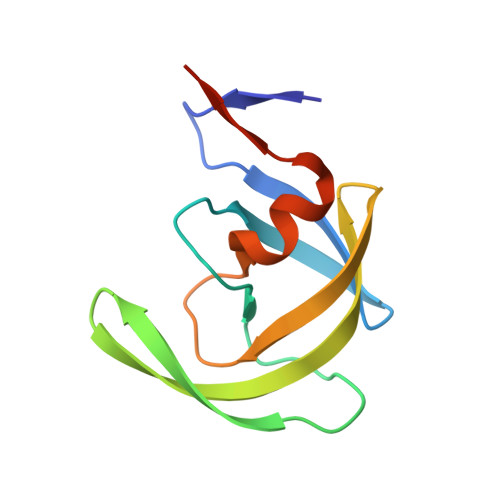
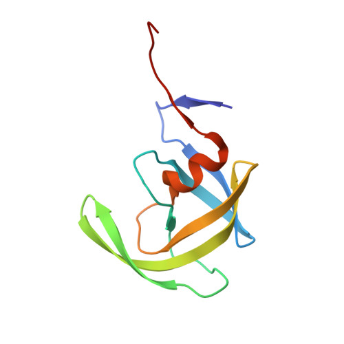
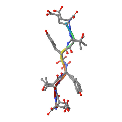
X-ray snapshot of HIV-1 protease in action: observation of tetrahedral intermediate and short ionic hydrogen bond SIHB with catalytic aspartate.

Das, A.Mahale, S.Prashar, V.Bihani, S.Ferrer, J.L.Hosur, M.V.

(2010) J Am Chem Soc 132: 6366-6373

  • DOI: https://doi.org/10.1021/ja100002b
  • Primary Citation of Related Structures:  
    5YRS

  • PubMed Abstract: 

    Structural snapshots of each step in the catalytic cycle would help development of inhibitors of human immunodeficiency virus type 1 protease (HIV-1 PR) as effective drugs against HIV/AIDS. We report here one snapshot obtained by determining the structure of enzyme-substrate complex under conditions where the catalytic activity of the enzyme is greatly reduced. The 1.76 A crystal structure shows the oligopeptide substrate, AETFYVDGAA, converted in situ into a gem-diol tetrahedral intermediate (TI). The gem-diol intermediate is neutral and one of the hydroxyl oxygens forms a very short hydrogen bond (2.2 A) with the anionic aspartate of the catalytic dyad, which is monoprotonated. Further, there is no hydrogen atom on the outer oxygen of the neutral aspartate. These two observations provide direct evidence that, in the reaction mechanism, hydrogen bonding between catalytic aspartate and scissile carbonyl oxygen facilitates water attack on the scissile carbon atom. Comparison with the structural snapshot of the biproduct complex involving the same substrate reveals the reorganization of the hydrogen bonds at the catalytic center as the enzymatic reaction progresses toward completion. Accumulation of TI in the crystals provides direct evidence that collapse of TI is the rate-limiting step of hydrolysis.


  • Organizational Affiliation

    Protein Crystallography Section, Solid State Physics Division, Bhabha Atomic Research Centre, Trombay, Mumbai-400085, India.


Macromolecules
Find similar proteins by:  (by identity cutoff)  |  3D Structure
Entity ID: 1
MoleculeChains Sequence LengthOrganismDetailsImage
PROTEASE104HIV-1 M:B_HXB2RMutation(s): 1 
Gene Names: gag-polHIV-1 PR B
EC: 3.4.23.16
UniProt
Find proteins for P04585 (Human immunodeficiency virus type 1 group M subtype B (isolate HXB2))
Explore P04585 
Go to UniProtKB:  P04585
Entity Groups  
Sequence Clusters30% Identity50% Identity70% Identity90% Identity95% Identity100% Identity
UniProt GroupP04585
Sequence Annotations
Expand
  • Reference Sequence
Find similar proteins by:  (by identity cutoff)  |  3D Structure
Entity ID: 2
MoleculeChains Sequence LengthOrganismDetailsImage
PROTEASE104HIV-1 M:B_HXB2RMutation(s): 1 
Gene Names: gag-polHIV-1 PR B
EC: 3.4.23.16
UniProt
Find proteins for P04585 (Human immunodeficiency virus type 1 group M subtype B (isolate HXB2))
Explore P04585 
Go to UniProtKB:  P04585
Entity Groups  
Sequence Clusters30% Identity50% Identity70% Identity90% Identity95% Identity100% Identity
UniProt GroupP04585
Sequence Annotations
Expand
  • Reference Sequence

Find similar proteins by:  Sequence   |   3D Structure  

Entity ID: 3
MoleculeChains Sequence LengthOrganismDetailsImage
RT-RH oligopeprtideC [auth X]6HIV-1 M:B_HXB2RMutation(s): 0 
UniProt
Find proteins for P04585 (Human immunodeficiency virus type 1 group M subtype B (isolate HXB2))
Explore P04585 
Go to UniProtKB:  P04585
Entity Groups  
UniProt GroupP04585
Sequence Annotations
Expand
  • Reference Sequence
Small Molecules
Modified Residues  1 Unique
IDChains TypeFormula2D DiagramParent
HPH
Query on HPH
C [auth X]PEPTIDE-LIKEC9 H13 N O2PHE
Experimental Data & Validation

Experimental Data

  • Method: X-RAY DIFFRACTION
  • Resolution: 1.76 Å
  • R-Value Free: 0.250 
  • R-Value Work: 0.223 
  • R-Value Observed: 0.223 
  • Space Group: P 61
Unit Cell:
Length ( Å )Angle ( ˚ )
a = 62.56α = 90
b = 62.56β = 90
c = 81.86γ = 120
Software Package:
Software NamePurpose
CNSrefinement
XDSdata reduction
XDSdata scaling
CNSphasing

Structure Validation

View Full Validation Report


Entry History & Funding Information

Deposition Data


Funding OrganizationLocationGrant Number
DAEIndiaInternal

Revision History  (Full details and data files)

  • Version 1.0: 2018-03-07
    Type: Initial release
  • Version 1.1: 2023-11-22
    Changes: Data collection, Database references, Derived calculations, Refinement description