5YLV

Crystal structure of the gastric proton pump complexed with SCH28080


Experimental Data Snapshot

  • Method: X-RAY DIFFRACTION
  • Resolution: 2.80 Å
  • R-Value Free: 0.292 
  • R-Value Work: 0.240 
  • R-Value Observed: 0.242 

wwPDB Validation   3D Report Full Report


Ligand Structure Quality Assessment 


This is version 1.3 of the entry. See complete history


Literature

Crystal structures of the gastric proton pump

Abe, K.Irie, K.Nakanishi, H.Suzuki, H.Fujiyoshi, Y.

(2018) Nature 556: 214-218

  • DOI: https://doi.org/10.1038/s41586-018-0003-8
  • Primary Citation of Related Structures:  
    5YLU, 5YLV

  • PubMed Abstract: 

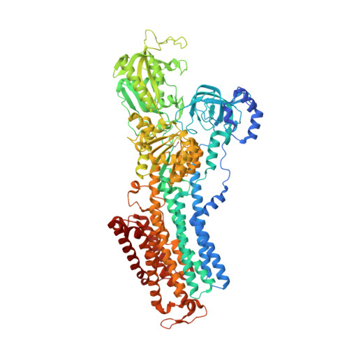
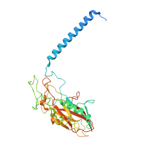
    The gastric proton pump-the H + , K + -ATPase-is a P-type ATPase responsible for acidifying the gastric juice down to pH 1. This corresponds to a million-fold proton gradient across the membrane of the parietal cell, the steepest known cation gradient of any mammalian tissue. The H + , K + -ATPase is an important target for drugs that treat gastric acid-related diseases. Here we present crystal structures of the H + , K + -ATPase in complex with two blockers, vonoprazan and SCH28080, in the luminal-open state, at 2.8 Å resolution. The drugs have partially overlapping but clearly distinct binding modes in the middle of a conduit running from the gastric lumen to the cation-binding site. The crystal structures suggest that the tight configuration at the cation-binding site lowers the pK a value of Glu820 sufficiently to enable the release of a proton even into the pH 1 environment of the stomach.


  • Organizational Affiliation

    Cellular and Structural Physiology Institute, Nagoya University, Nagoya, Japan. kabe@cespi.nagoya-u.ac.jp.


Macromolecules
Find similar proteins by:  (by identity cutoff)  |  3D Structure
Entity ID: 1
MoleculeChains Sequence LengthOrganismDetailsImage
Potassium-transporting ATPase alpha chain 11,034Sus scrofaMutation(s): 3 
Gene Names: ATP4A
EC: 3.6.3.10
Membrane Entity: Yes 
UniProt
Find proteins for P19156 (Sus scrofa)
Explore P19156 
Go to UniProtKB:  P19156
Entity Groups  
Sequence Clusters30% Identity50% Identity70% Identity90% Identity95% Identity100% Identity
UniProt GroupP19156
Sequence Annotations
Expand
  • Reference Sequence
Find similar proteins by:  (by identity cutoff)  |  3D Structure
Entity ID: 2
MoleculeChains Sequence LengthOrganismDetailsImage
Potassium-transporting ATPase subunit beta289Sus scrofaMutation(s): 0 
Gene Names: ATP4B
Membrane Entity: Yes 
UniProt
Find proteins for P18434 (Sus scrofa)
Explore P18434 
Go to UniProtKB:  P18434
Entity Groups  
Sequence Clusters30% Identity50% Identity70% Identity90% Identity95% Identity100% Identity
UniProt GroupP18434
Sequence Annotations
Expand
  • Reference Sequence
Small Molecules
Ligands 6 Unique
IDChains Name / Formula / InChI Key2D Diagram3D Interactions
PCW
Query on PCW

Download Ideal Coordinates CCD File 
L [auth B]1,2-DIOLEOYL-SN-GLYCERO-3-PHOSPHOCHOLINE
C44 H85 N O8 P
SNKAWJBJQDLSFF-NVKMUCNASA-O
CE1
Query on CE1

Download Ideal Coordinates CCD File 
E [auth A],
M [auth B]
O-DODECANYL OCTAETHYLENE GLYCOL
C28 H58 O9
YYELLDKEOUKVIQ-UHFFFAOYSA-N
8WX
Query on 8WX

Download Ideal Coordinates CCD File 
C [auth A]2-(2-methyl-8-phenylmethoxy-imidazo[1,2-a]pyridin-3-yl)ethanenitrile
C17 H15 N3 O
PYKJFEPAUKAXNN-UHFFFAOYSA-N
NAG
Query on NAG

Download Ideal Coordinates CCD File 
I [auth B],
J [auth B],
K [auth B]
2-acetamido-2-deoxy-beta-D-glucopyranose
C8 H15 N O6
OVRNDRQMDRJTHS-FMDGEEDCSA-N
RB
Query on RB

Download Ideal Coordinates CCD File 
F [auth A],
G [auth A],
H [auth A]
RUBIDIUM ION
Rb
NCCSSGKUIKYAJD-UHFFFAOYSA-N
MG
Query on MG

Download Ideal Coordinates CCD File 
D [auth A]MAGNESIUM ION
Mg
JLVVSXFLKOJNIY-UHFFFAOYSA-N
Modified Residues  1 Unique
IDChains TypeFormula2D DiagramParent
BFD
Query on BFD
A
L-PEPTIDE LINKINGC4 H6 Be F3 N O4ASP
Binding Affinity Annotations 
IDSourceBinding Affinity
8WX BindingDB:  5YLV IC50: min: 65, max: 1590 (nM) from 3 assay(s)
Experimental Data & Validation

Experimental Data

  • Method: X-RAY DIFFRACTION
  • Resolution: 2.80 Å
  • R-Value Free: 0.292 
  • R-Value Work: 0.240 
  • R-Value Observed: 0.242 
  • Space Group: P 31 2 1
Unit Cell:
Length ( Å )Angle ( ˚ )
a = 105.05α = 90
b = 105.05β = 90
c = 368.54γ = 120
Software Package:
Software NamePurpose
PHENIXrefinement
XDSdata reduction
XDSdata scaling
PHASERphasing

Structure Validation

View Full Validation Report


Ligand Structure Quality Assessment 


Entry History 

Deposition Data

Revision History  (Full details and data files)

  • Version 1.0: 2018-04-11
    Type: Initial release
  • Version 1.1: 2018-04-18
    Changes: Data collection, Database references
  • Version 1.2: 2020-07-29
    Type: Remediation
    Reason: Carbohydrate remediation
    Changes: Data collection, Derived calculations, Structure summary
  • Version 1.3: 2023-11-22
    Changes: Data collection, Database references, Refinement description, Structure summary