5WXK

EarP bound with domain I of EF-P


Experimental Data Snapshot

  • Method: X-RAY DIFFRACTION
  • Resolution: 1.80 Å
  • R-Value Free: 0.202 
  • R-Value Work: 0.157 
  • R-Value Observed: 0.159 

wwPDB Validation   3D Report Full Report


Ligand Structure Quality Assessment 


This is version 1.2 of the entry. See complete history


Literature

Structural basis of protein arginine rhamnosylation by glycosyltransferase EarP

Sengoku, T.Suzuki, T.Dohmae, N.Watanabe, C.Honma, T.Hikida, Y.Yamaguchi, Y.Takahashi, H.Yokoyama, S.Yanagisawa, T.

(2018) Nat Chem Biol 14: 368-374

  • DOI: https://doi.org/10.1038/s41589-018-0002-y
  • Primary Citation of Related Structures:  
    5WXI, 5WXJ, 5WXK, 5XVR

  • PubMed Abstract: 

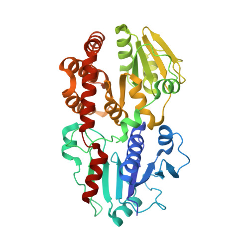
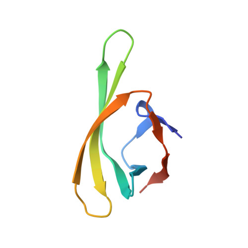
    Protein glycosylation regulates many cellular processes. Numerous glycosyltransferases with broad substrate specificities have been structurally characterized. A novel inverting glycosyltransferase, EarP, specifically transfers rhamnose from dTDP-β-L-rhamnose to Arg32 of bacterial translation elongation factor P (EF-P) to activate its function. Here we report a crystallographic study of Neisseria meningitidis EarP. The EarP structure contains two tandem Rossmann-fold domains, which classifies EarP in glycosyltransferase superfamily B. In contrast to other structurally characterized protein glycosyltransferases, EarP binds the entire β-sheet structure of EF-P domain I through numerous interactions that specifically recognize its conserved residues. Thus Arg32 is properly located at the active site, and causes structural change in a conserved dTDP-β-L-rhamnose-binding loop of EarP. Rhamnosylation by EarP should occur via an S N 2 reaction, with Asp20 as the general base. The Arg32 binding and accompanying structural change of EarP may induce a change in the rhamnose-ring conformation suitable for the reaction.


  • Organizational Affiliation

    RIKEN Structural Biology Laboratory, Yokohama, Japan.


Macromolecules
Find similar proteins by:  (by identity cutoff)  |  3D Structure
Entity ID: 1
MoleculeChains Sequence LengthOrganismDetailsImage
EarP382Neisseria meningitidis H44/76Mutation(s): 0 
Gene Names: NMH_0797
UniProt
Find proteins for E6MVV9 (Neisseria meningitidis serogroup B / serotype 15 (strain H44/76))
Explore E6MVV9 
Go to UniProtKB:  E6MVV9
Entity Groups  
Sequence Clusters30% Identity50% Identity70% Identity90% Identity95% Identity100% Identity
UniProt GroupE6MVV9
Sequence Annotations
Expand
  • Reference Sequence
Find similar proteins by:  (by identity cutoff)  |  3D Structure
Entity ID: 2
MoleculeChains Sequence LengthOrganismDetailsImage
Elongation factor P70Neisseria meningitidis H44/76Mutation(s): 0 
Gene Names: efpNMH_0798
UniProt
Find proteins for E6MVW0 (Neisseria meningitidis serogroup B / serotype 15 (strain H44/76))
Explore E6MVW0 
Go to UniProtKB:  E6MVW0
Entity Groups  
Sequence Clusters30% Identity50% Identity70% Identity90% Identity95% Identity100% Identity
UniProt GroupE6MVW0
Sequence Annotations
Expand
  • Reference Sequence
Small Molecules
Ligands 5 Unique
IDChains Name / Formula / InChI Key2D Diagram3D Interactions
TYD
Query on TYD

Download Ideal Coordinates CCD File 
C [auth A]THYMIDINE-5'-DIPHOSPHATE
C10 H16 N2 O11 P2
UJLXYODCHAELLY-XLPZGREQSA-N
SO4
Query on SO4

Download Ideal Coordinates CCD File 
D [auth A],
E [auth A],
F [auth A],
G [auth A]
SULFATE ION
O4 S
QAOWNCQODCNURD-UHFFFAOYSA-L
GOL
Query on GOL

Download Ideal Coordinates CCD File 
K [auth A],
M [auth B]
GLYCEROL
C3 H8 O3
PEDCQBHIVMGVHV-UHFFFAOYSA-N
BME
Query on BME

Download Ideal Coordinates CCD File 
H [auth A],
I [auth A],
J [auth A]
BETA-MERCAPTOETHANOL
C2 H6 O S
DGVVWUTYPXICAM-UHFFFAOYSA-N
NI
Query on NI

Download Ideal Coordinates CCD File 
L [auth A]NICKEL (II) ION
Ni
VEQPNABPJHWNSG-UHFFFAOYSA-N
Experimental Data & Validation

Experimental Data

  • Method: X-RAY DIFFRACTION
  • Resolution: 1.80 Å
  • R-Value Free: 0.202 
  • R-Value Work: 0.157 
  • R-Value Observed: 0.159 
  • Space Group: C 1 2 1
Unit Cell:
Length ( Å )Angle ( ˚ )
a = 122.204α = 90
b = 57.254β = 101.44
c = 72.831γ = 90
Software Package:
Software NamePurpose
PHENIXrefinement
XDSdata extraction
PHENIXphasing

Structure Validation

View Full Validation Report


Ligand Structure Quality Assessment 


Entry History & Funding Information

Deposition Data


Funding OrganizationLocationGrant Number
MEXTJapan--
AMEDJapan--

Revision History  (Full details and data files)

  • Version 1.0: 2018-02-28
    Type: Initial release
  • Version 1.1: 2018-04-04
    Changes: Data collection, Database references
  • Version 1.2: 2024-04-03
    Changes: Data collection, Database references, Derived calculations, Refinement description