5WN9

Structure of antibody 2D10 bound to the central conserved region of RSV G


Experimental Data Snapshot

  • Method: X-RAY DIFFRACTION
  • Resolution: 1.55 Å
  • R-Value Free: 0.185 
  • R-Value Work: 0.169 
  • R-Value Observed: 0.170 

wwPDB Validation   3D Report Full Report


This is version 1.2 of the entry. See complete history


Literature

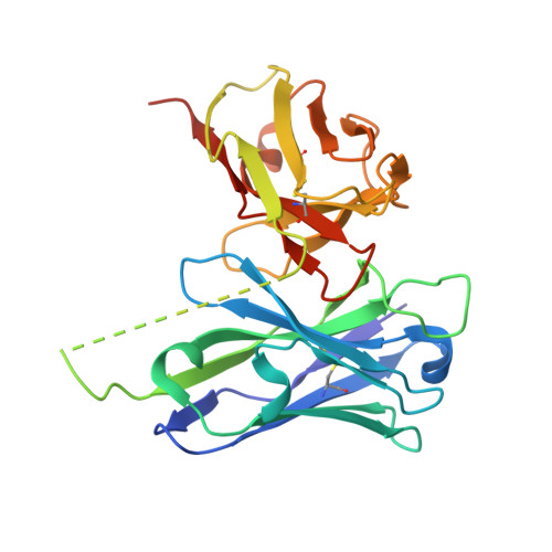
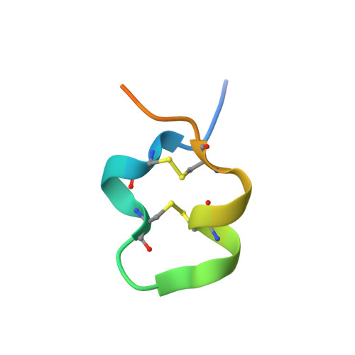
Structures of respiratory syncytial virus G antigen bound to broadly neutralizing antibodies.

Fedechkin, S.O.George, N.L.Wolff, J.T.Kauvar, L.M.DuBois, R.M.

(2018) Sci Immunol 3

  • DOI: https://doi.org/10.1126/sciimmunol.aar3534
  • Primary Citation of Related Structures:  
    5WN9, 5WNA, 5WNB

  • PubMed Abstract: 

    Respiratory syncytial virus (RSV) is a top cause of severe lower respiratory tract disease and mortality in young children and the elderly. The viral envelope G glycoprotein contributes to pathogenesis through its roles in host cell attachment and modulation of host immunity. Although the G glycoprotein is a target of protective RSV-neutralizing antibodies, its development as a vaccine antigen has been hindered by its heterogeneous glycosylation and sequence variability outside a conserved central domain (CCD). We describe the cocrystal structures of two high-affinity broadly neutralizing human monoclonal antibodies bound to the RSV G CCD. The antibodies bind to neighboring conformational epitopes, which we named antigenic sites γ1 and γ2, that span a highly conserved surface, illuminating an important region of vulnerability. We further show that isolated RSV G CCD activates the chemokine receptor CX3CR1 and that antibodies block this activity. These studies provide a template for rational vaccine design targeting this key contributor to RSV disease.


  • Organizational Affiliation

    Department of Biomolecular Engineering, University of California, Santa Cruz, Santa Cruz, CA 95064, USA.


Macromolecules
Find similar proteins by:  (by identity cutoff)  |  3D Structure
Entity ID: 1
MoleculeChains Sequence LengthOrganismDetailsImage
scFv 2D10A [auth H]260Homo sapiensMutation(s): 0 
Entity Groups  
Sequence Clusters30% Identity50% Identity70% Identity90% Identity95% Identity100% Identity
Sequence Annotations
Expand
  • Reference Sequence
Find similar proteins by:  (by identity cutoff)  |  3D Structure
Entity ID: 2
MoleculeChains Sequence LengthOrganismDetailsImage
Major surface glycoprotein GB [auth A]29Human respiratory syncytial virus A2Mutation(s): 0 
UniProt
Find proteins for P03423 (Human respiratory syncytial virus A (strain A2))
Explore P03423 
Go to UniProtKB:  P03423
Entity Groups  
Sequence Clusters30% Identity50% Identity70% Identity90% Identity95% Identity100% Identity
UniProt GroupP03423
Sequence Annotations
Expand
  • Reference Sequence
Experimental Data & Validation

Experimental Data

  • Method: X-RAY DIFFRACTION
  • Resolution: 1.55 Å
  • R-Value Free: 0.185 
  • R-Value Work: 0.169 
  • R-Value Observed: 0.170 
  • Space Group: P 21 21 21
Unit Cell:
Length ( Å )Angle ( ˚ )
a = 44.843α = 90
b = 56.387β = 90
c = 126.148γ = 90
Software Package:
Software NamePurpose
PHENIXrefinement
HKL-2000data reduction
SCALEPACKdata scaling
PDB_EXTRACTdata extraction
Cootmodel building
PHASERphasing

Structure Validation

View Full Validation Report


Entry History 

Deposition Data

Revision History  (Full details and data files)

  • Version 1.0: 2018-03-14
    Type: Initial release
  • Version 1.1: 2018-03-21
    Changes: Database references
  • Version 1.2: 2023-10-04
    Changes: Data collection, Database references, Refinement description