5W5Z

Crystal structure of BAXP168G in complex with an activating antibody at high resolution


Experimental Data Snapshot

  • Method: X-RAY DIFFRACTION
  • Resolution: 2.00 Å
  • R-Value Free: 0.268 
  • R-Value Work: 0.217 
  • R-Value Observed: 0.219 

wwPDB Validation   3D Report Full Report


This is version 2.0 of the entry. See complete history


Literature

Ensemble Properties of Bax Determine Its Function.

Robin, A.Y.Iyer, S.Birkinshaw, R.W.Sandow, J.Wardak, A.Luo, C.S.Shi, M.Webb, A.I.Czabotar, P.E.Kluck, R.M.Colman, P.M.

(2018) Structure 26: 1346

  • DOI: https://doi.org/10.1016/j.str.2018.07.006
  • Primary Citation of Related Structures:  
    5W5X, 5W5Z, 5W60, 5W61, 5W62, 5W63

  • PubMed Abstract: 

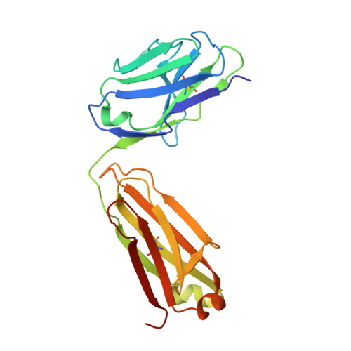
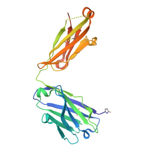
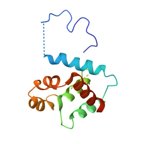
    BAX and BAK are essential mediators of intrinsic apoptosis that permeabilize the mitochondrial outer membrane. BAX activation requires its translocation from cytosol to mitochondria where conformational changes cause its oligomerization. To better understand the critical step of translocation, we examined its blockade by mutation near the C terminus (P168G) or by antibody binding near the N terminus. Similarities in the crystal structures of wild-type and BAX P168G but significant other differences suggest that cytosolic BAX exists as an ensemble of conformers, and that the distribution of conformers within the ensemble determines the different functions of wild-type and mutant proteins. We also describe the structure of BAX in complex with an antibody, 3C10, that inhibits cytosolic BAX by limiting exposure of the membrane-associating helix α9, as does the P168G mutation. Our data for both means of BAX inhibition argue for an allosteric model of BAX regulation that derives from properties of the ensemble of conformers.


  • Organizational Affiliation

    Walter and Eliza Hall Institute of Medical Research, 1G Royal Parade, Parkville, VIC 3052, Australia; Department of Medical Biology, The University of Melbourne, Melbourne, VIC 3052, Australia.


Macromolecules
Find similar proteins by:  (by identity cutoff)  |  3D Structure
Entity ID: 1
MoleculeChains Sequence LengthOrganismDetailsImage
3C10 Fab light chainA [auth L]213Rattus norvegicusMutation(s): 0 
Entity Groups  
Sequence Clusters30% Identity50% Identity70% Identity90% Identity95% Identity100% Identity
Sequence Annotations
Expand
  • Reference Sequence
Find similar proteins by:  (by identity cutoff)  |  3D Structure
Entity ID: 2
MoleculeChains Sequence LengthOrganismDetailsImage
3C10 Fab heavy chainB [auth H]234Rattus norvegicusMutation(s): 0 
Entity Groups  
Sequence Clusters30% Identity50% Identity70% Identity90% Identity95% Identity100% Identity
Sequence Annotations
Expand
  • Reference Sequence
Find similar proteins by:  (by identity cutoff)  |  3D Structure
Entity ID: 3
MoleculeChains Sequence LengthOrganismDetailsImage
Apoptosis regulator BAXC [auth A]163Homo sapiensMutation(s): 3 
Gene Names: BAXBCL2L4
UniProt & NIH Common Fund Data Resources
Find proteins for Q07812 (Homo sapiens)
Explore Q07812 
Go to UniProtKB:  Q07812
PHAROS:  Q07812
GTEx:  ENSG00000087088 
Entity Groups  
Sequence Clusters30% Identity50% Identity70% Identity90% Identity95% Identity100% Identity
UniProt GroupQ07812
Sequence Annotations
Expand
  • Reference Sequence
Small Molecules
Ligands 4 Unique
IDChains Name / Formula / InChI Key2D Diagram3D Interactions
MES
Query on MES

Download Ideal Coordinates CCD File 
K [auth H]2-(N-MORPHOLINO)-ETHANESULFONIC ACID
C6 H13 N O4 S
SXGZJKUKBWWHRA-UHFFFAOYSA-N
PG4
Query on PG4

Download Ideal Coordinates CCD File 
G [auth L],
H [auth L]
TETRAETHYLENE GLYCOL
C8 H18 O5
UWHCKJMYHZGTIT-UHFFFAOYSA-N
PGE
Query on PGE

Download Ideal Coordinates CCD File 
I [auth H],
J [auth H]
TRIETHYLENE GLYCOL
C6 H14 O4
ZIBGPFATKBEMQZ-UHFFFAOYSA-N
EDO
Query on EDO

Download Ideal Coordinates CCD File 
D [auth L],
E [auth L],
F [auth L]
1,2-ETHANEDIOL
C2 H6 O2
LYCAIKOWRPUZTN-UHFFFAOYSA-N
Modified Residues  1 Unique
IDChains TypeFormula2D DiagramParent
PCA
Query on PCA
B [auth H]L-PEPTIDE LINKINGC5 H7 N O3GLN
Experimental Data & Validation

Experimental Data

  • Method: X-RAY DIFFRACTION
  • Resolution: 2.00 Å
  • R-Value Free: 0.268 
  • R-Value Work: 0.217 
  • R-Value Observed: 0.219 
  • Space Group: P 41 21 2
Unit Cell:
Length ( Å )Angle ( ˚ )
a = 67.863α = 90
b = 67.863β = 90
c = 264.243γ = 90
Software Package:
Software NamePurpose
PHENIXrefinement
XDSdata reduction
XSCALEdata scaling
PHASERphasing

Structure Validation

View Full Validation Report


Entry History 

Deposition Data

Revision History  (Full details and data files)

  • Version 1.0: 2018-06-27
    Type: Initial release
  • Version 1.1: 2018-08-29
    Changes: Data collection, Database references
  • Version 1.2: 2018-09-05
    Changes: Data collection, Database references
  • Version 1.3: 2018-10-17
    Changes: Data collection, Database references
  • Version 2.0: 2023-10-04
    Changes: Data collection, Database references, Polymer sequence, Refinement description