5W34

Crystal structure of the RNA polymerase domain (RPD) of Mycobacterium tuberculosis primase DnaG in complex with double-stranded DNA GACCGGAAGTGG


Experimental Data Snapshot

  • Method: X-RAY DIFFRACTION
  • Resolution: 2.95 Å
  • R-Value Free: 0.285 
  • R-Value Work: 0.236 
  • R-Value Observed: 0.238 

wwPDB Validation   3D Report Full Report


This is version 1.2 of the entry. See complete history


Literature

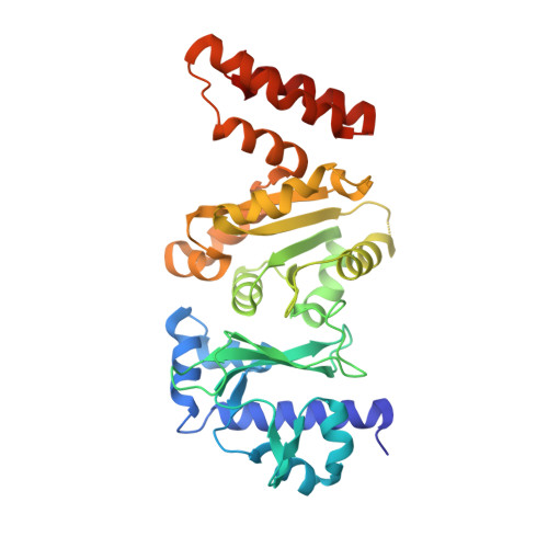
Structures of the Catalytic Domain of Bacterial Primase DnaG in Complexes with DNA Provide Insight into Key Priming Events.

Hou, C.Biswas, T.Tsodikov, O.V.

(2018) Biochemistry 57: 2084-2093

  • DOI: https://doi.org/10.1021/acs.biochem.8b00036
  • Primary Citation of Related Structures:  
    5W33, 5W34, 5W35, 5W36

  • PubMed Abstract: 

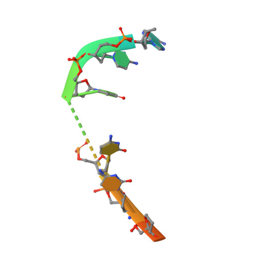
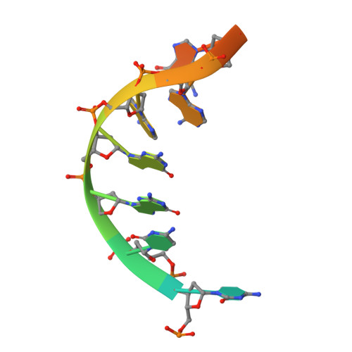
    Bacterial primase DnaG is an essential nucleic acid polymerase that generates primers for replication of chromosomal DNA. The mechanism of DnaG remains unclear due to the paucity of structural information on DnaG in complexes with other replisome components. Here we report the first crystal structures of noncovalent DnaG-DNA complexes, obtained with the RNA polymerase domain of Mycobacterium tuberculosis DnaG and various DNA ligands. One structure, obtained with ds DNA, reveals interactions with DnaG as it slides on ds DNA and suggests how DnaG binds template for primer synthesis. In another structure, DNA in the active site of DnaG mimics the primer, providing insight into mechanisms for the nucleotide transfer and DNA translocation. In conjunction with the recent cryo-EM structure of the bacteriophage T7 replisome, this study yields a model for primer elongation and hand-off to DNA polymerase.


  • Organizational Affiliation

    Department of Pharmaceutical Sciences, College of Pharmacy , University of Kentucky , Lexington , Kentucky 40536 , United States.


Macromolecules

Find similar proteins by:  (by identity cutoff)  |  3D Structure
Entity ID: 1
MoleculeChains Sequence LengthOrganismDetailsImage
DNA primase
A, B
325Mycobacterium tuberculosis H37RvMutation(s): 0 
Gene Names: dnaGRv2343cMTCY98.12c
EC: 2.7.7
UniProt
Find proteins for P9WNW1 (Mycobacterium tuberculosis (strain ATCC 25618 / H37Rv))
Explore P9WNW1 
Go to UniProtKB:  P9WNW1
Entity Groups  
Sequence Clusters30% Identity50% Identity70% Identity90% Identity95% Identity100% Identity
UniProt GroupP9WNW1
Sequence Annotations
Expand
  • Reference Sequence

Find similar nucleic acids by:  Sequence   |   3D Structure  

Entity ID: 2
MoleculeChains LengthOrganismImage
DNA oligomer 5'-CCACTTCCGGTC12synthetic construct
Sequence Annotations
Expand
  • Reference Sequence

Find similar nucleic acids by:  Sequence   |   3D Structure  

Entity ID: 3
MoleculeChains LengthOrganismImage
DNA oligomer 5'-GACCGGAAGTGG12synthetic construct
Sequence Annotations
Expand
  • Reference Sequence
Experimental Data & Validation

Experimental Data

  • Method: X-RAY DIFFRACTION
  • Resolution: 2.95 Å
  • R-Value Free: 0.285 
  • R-Value Work: 0.236 
  • R-Value Observed: 0.238 
  • Space Group: P 1 21 1
Unit Cell:
Length ( Å )Angle ( ˚ )
a = 43.781α = 90
b = 212.116β = 107.87
c = 47.418γ = 90
Software Package:
Software NamePurpose
REFMACrefinement
HKL-2000data reduction
HKL-2000data scaling
PHASERphasing

Structure Validation

View Full Validation Report


Entry History 

Deposition Data

Revision History  (Full details and data files)

  • Version 1.0: 2018-03-28
    Type: Initial release
  • Version 1.1: 2018-04-18
    Changes: Data collection, Database references
  • Version 1.2: 2023-10-04
    Changes: Data collection, Database references, Refinement description