5VQA

Structure of human TRIP13, ATP-bound form


Experimental Data Snapshot

  • Method: X-RAY DIFFRACTION
  • Resolution: 2.54 Å
  • R-Value Free: 0.241 
  • R-Value Work: 0.197 
  • R-Value Observed: 0.199 

wwPDB Validation   3D Report Full Report


Ligand Structure Quality Assessment 


This is version 1.3 of the entry. See complete history


Literature

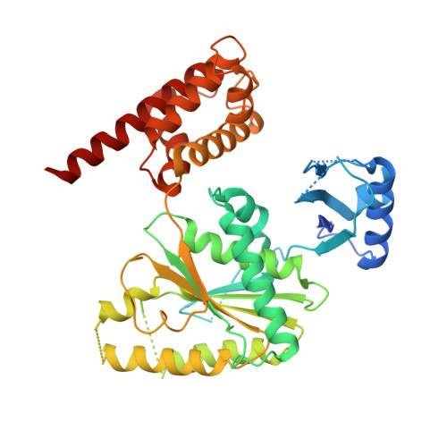
The AAA+ ATPase TRIP13 remodels HORMA domains through N-terminal engagement and unfolding.

Ye, Q.Kim, D.H.Dereli, I.Rosenberg, S.C.Hagemann, G.Herzog, F.Toth, A.Cleveland, D.W.Corbett, K.D.

(2017) EMBO J 36: 2419-2434

  • DOI: https://doi.org/10.15252/embj.201797291
  • Primary Citation of Related Structures:  
    5VQ9, 5VQA

  • PubMed Abstract: 

    Proteins of the conserved HORMA domain family, including the spindle assembly checkpoint protein MAD2 and the meiotic HORMADs, assemble into signaling complexes by binding short peptides termed "closure motifs". The AAA+ ATPase TRIP13 regulates both MAD2 and meiotic HORMADs by disassembling these HORMA domain-closure motif complexes, but its mechanisms of substrate recognition and remodeling are unknown. Here, we combine X-ray crystallography and crosslinking mass spectrometry to outline how TRIP13 recognizes MAD2 with the help of the adapter protein p31 comet We show that p31 comet binding to the TRIP13 N-terminal domain positions the disordered MAD2 N-terminus for engagement by the TRIP13 "pore loops", which then unfold MAD2 in the presence of ATP N-terminal truncation of MAD2 renders it refractory to TRIP13 action in vitro , and in cells causes spindle assembly checkpoint defects consistent with loss of TRIP13 function. Similar truncation of HORMAD1 in mouse spermatocytes compromises its TRIP13-mediated removal from meiotic chromosomes, highlighting a conserved mechanism for recognition and disassembly of HORMA domain-closure motif complexes by TRIP13.


  • Organizational Affiliation

    Ludwig Institute for Cancer Research, San Diego Branch, La Jolla, CA, USA.


Macromolecules
Find similar proteins by:  (by identity cutoff)  |  3D Structure
Entity ID: 1
MoleculeChains Sequence LengthOrganismDetailsImage
Pachytene checkpoint protein 2 homolog432Homo sapiensMutation(s): 1 
Gene Names: TRIP13PCH2
UniProt & NIH Common Fund Data Resources
Find proteins for Q15645 (Homo sapiens)
Explore Q15645 
Go to UniProtKB:  Q15645
PHAROS:  Q15645
GTEx:  ENSG00000071539 
Entity Groups  
Sequence Clusters30% Identity50% Identity70% Identity90% Identity95% Identity100% Identity
UniProt GroupQ15645
Sequence Annotations
Expand
  • Reference Sequence
Small Molecules
Ligands 1 Unique
IDChains Name / Formula / InChI Key2D Diagram3D Interactions
ATP
Query on ATP

Download Ideal Coordinates CCD File 
B [auth A]ADENOSINE-5'-TRIPHOSPHATE
C10 H16 N5 O13 P3
ZKHQWZAMYRWXGA-KQYNXXCUSA-N
Experimental Data & Validation

Experimental Data

Unit Cell:
Length ( Å )Angle ( ˚ )
a = 98.479α = 90
b = 98.479β = 90
c = 120.979γ = 120
Software Package:
Software NamePurpose
PHENIXrefinement
XDSdata reduction
Aimlessdata scaling
PHASERphasing

Structure Validation

View Full Validation Report


Ligand Structure Quality Assessment 


Entry History 

Deposition Data

Revision History  (Full details and data files)

  • Version 1.0: 2017-06-14
    Type: Initial release
  • Version 1.1: 2017-07-12
    Changes: Database references
  • Version 1.2: 2017-08-30
    Changes: Database references
  • Version 1.3: 2023-10-04
    Changes: Author supporting evidence, Data collection, Database references, Refinement description