5VPH

CRYSTAL STRUCTURE OF DER P 1 COMPLEXED WITH FAB 4C1


Experimental Data Snapshot

  • Method: X-RAY DIFFRACTION
  • Resolution: 2.50 Å
  • R-Value Free: 0.229 
  • R-Value Work: 0.178 
  • R-Value Observed: 0.180 

wwPDB Validation   3D Report Full Report


This is version 1.4 of the entry. See complete history


Literature

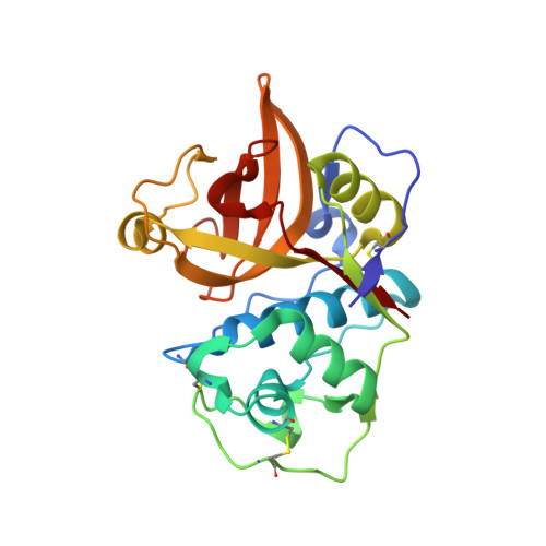
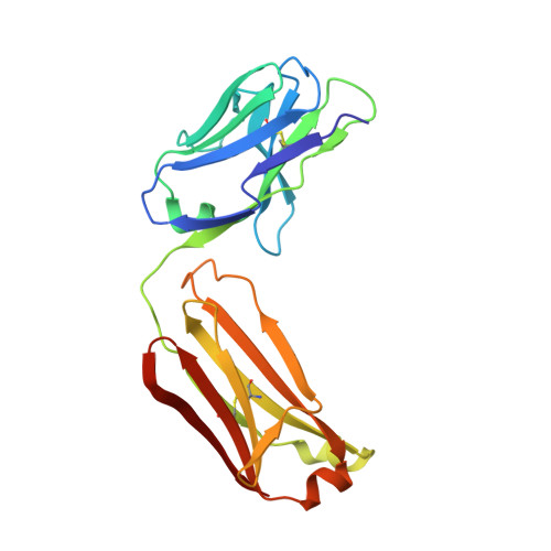
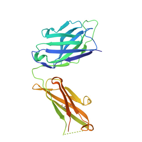
Molecular Determinants For Antibody Binding On Group 1 House Dust Mite Allergens.

Chruszcz, M.Pomes, A.Glesner, J.Vailes, L.D.Osinski, T.Porebski, P.J.Majorek, K.A.Heymann, P.W.Platts-Mills, T.A.Minor, W.Chapman, M.D.

(2012) J Biol Chem 287: 7388

  • DOI: https://doi.org/10.1074/jbc.M111.311159
  • Primary Citation of Related Structures:  
    3RVT, 3RVU, 5VPG, 5VPH, 5VPL

  • PubMed Abstract: 

    House dust mites produce potent allergens, Der p 1 and Der f 1, that cause allergic sensitization and asthma. Der p 1 and Der f 1 are cysteine proteases that elicit IgE responses in 80% of mite-allergic subjects and have proinflammatory properties. Their antigenic structure is unknown. Here, we present crystal structures of natural Der p 1 and Der f 1 in complex with a monoclonal antibody, 4C1, which binds to a unique cross-reactive epitope on both allergens associated with IgE recognition. The 4C1 epitope is formed by almost identical amino acid sequences and contact residues. Mutations of the contact residues abrogate mAb 4C1 binding and reduce IgE antibody binding. These surface-exposed residues are molecular targets that can be exploited for development of recombinant allergen vaccines.


  • Organizational Affiliation

    Department of Molecular Physiology and Biological Physics, University of Virginia, Charlottesville, Virginia 22908, USA.


Macromolecules
Find similar proteins by:  (by identity cutoff)  |  3D Structure
Entity ID: 1
MoleculeChains Sequence LengthOrganismDetailsImage
Der p 1 allergen222Dermatophagoides pteronyssinusMutation(s): 0 
UniProt
Find proteins for P08176 (Dermatophagoides pteronyssinus)
Explore P08176 
Go to UniProtKB:  P08176
Entity Groups  
Sequence Clusters30% Identity50% Identity70% Identity90% Identity95% Identity100% Identity
UniProt GroupP08176
Sequence Annotations
Expand
  • Reference Sequence
Find similar proteins by:  (by identity cutoff)  |  3D Structure
Entity ID: 2
MoleculeChains Sequence LengthOrganismDetailsImage
FAB 4C1 - LIGHT CHAINB [auth C]213Mus musculusMutation(s): 0 
Entity Groups  
Sequence Clusters30% Identity50% Identity70% Identity90% Identity95% Identity100% Identity
Sequence Annotations
Expand
  • Reference Sequence
Find similar proteins by:  (by identity cutoff)  |  3D Structure
Entity ID: 3
MoleculeChains Sequence LengthOrganismDetailsImage
FAB 4C1 - HEAVY CHAINC [auth D]255Mus musculusMutation(s): 0 
Entity Groups  
Sequence Clusters30% Identity50% Identity70% Identity90% Identity95% Identity100% Identity
Sequence Annotations
Expand
  • Reference Sequence
Small Molecules
Experimental Data & Validation

Experimental Data

  • Method: X-RAY DIFFRACTION
  • Resolution: 2.50 Å
  • R-Value Free: 0.229 
  • R-Value Work: 0.178 
  • R-Value Observed: 0.180 
  • Space Group: P 21 21 21
Unit Cell:
Length ( Å )Angle ( ˚ )
a = 50.109α = 90
b = 59.377β = 90
c = 236.967γ = 90
Software Package:
Software NamePurpose
REFMACrefinement
HKL-3000data reduction
HKL-3000data scaling
MOLREPphasing
HKL-3000phasing
Cootmodel building

Structure Validation

View Full Validation Report


Entry History 

Deposition Data

Revision History  (Full details and data files)

  • Version 1.0: 2017-05-24
    Type: Initial release
  • Version 1.1: 2017-08-02
    Changes: Database references, Derived calculations, Source and taxonomy, Structure summary
  • Version 1.2: 2017-11-22
    Changes: Refinement description
  • Version 1.3: 2022-04-13
    Changes: Database references, Structure summary
  • Version 1.4: 2023-10-04
    Changes: Data collection, Refinement description