5VGD

Crystal Structure of HLA-C*0501 in complex with SAE


Experimental Data Snapshot

  • Method: X-RAY DIFFRACTION
  • Resolution: 2.32 Å
  • R-Value Free: 0.297 
  • R-Value Work: 0.220 
  • R-Value Observed: 0.224 

wwPDB Validation   3D Report Full Report


This is version 1.1 of the entry. See complete history


Literature

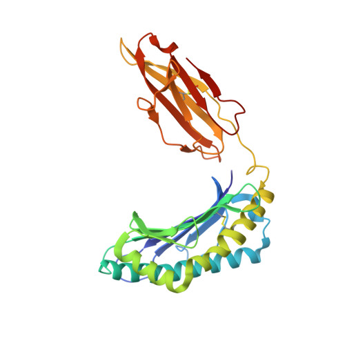
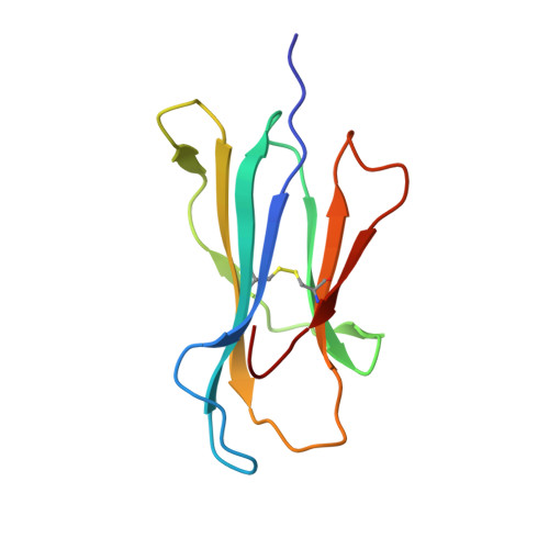
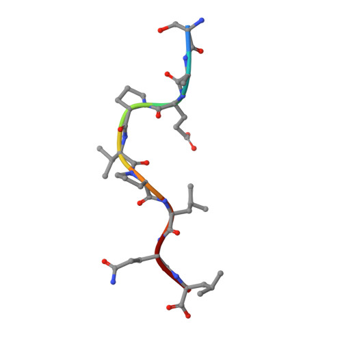
Structural and regulatory diversity shape HLA-C protein expression levels.

Kaur, G.Gras, S.Mobbs, J.I.Vivian, J.P.Cortes, A.Barber, T.Kuttikkatte, S.B.Jensen, L.T.Attfield, K.E.Dendrou, C.A.Carrington, M.McVean, G.Purcell, A.W.Rossjohn, J.Fugger, L.

(2017) Nat Commun 8: 15924-15924

  • DOI: https://doi.org/10.1038/ncomms15924
  • Primary Citation of Related Structures:  
    5VGD, 5VGE

  • PubMed Abstract: 

    Expression of HLA-C varies widely across individuals in an allele-specific manner. This variation in expression can influence efficacy of the immune response, as shown for infectious and autoimmune diseases. MicroRNA binding partially influences differential HLA-C expression, but the additional contributing factors have remained undetermined. Here we use functional and structural analyses to demonstrate that HLA-C expression is modulated not just at the RNA level, but also at the protein level. Specifically, we show that variation in exons 2 and 3, which encode the α1/α2 domains, drives differential expression of HLA-C allomorphs at the cell surface by influencing the structure of the peptide-binding cleft and the diversity of peptides bound by the HLA-C molecules. Together with a phylogenetic analysis, these results highlight the diversity and long-term balancing selection of regulatory factors that modulate HLA-C expression.


  • Organizational Affiliation

    MRC Human Immunology Unit, Weatherall Institute of Molecular Medicine, John Radcliffe Hospital, University of Oxford, Oxford OX3 9DS, UK.


Macromolecules
Find similar proteins by:  (by identity cutoff)  |  3D Structure
Entity ID: 1
MoleculeChains Sequence LengthOrganismDetailsImage
HLA class I histocompatibility antigen, Cw-5 alpha chain277Homo sapiensMutation(s): 0 
Gene Names: HLA-CHLAC
UniProt & NIH Common Fund Data Resources
Find proteins for P10321 (Homo sapiens)
Explore P10321 
Go to UniProtKB:  P10321
PHAROS:  P10321
GTEx:  ENSG00000204525 
Entity Groups  
Sequence Clusters30% Identity50% Identity70% Identity90% Identity95% Identity100% Identity
UniProt GroupP10321
Sequence Annotations
Expand
  • Reference Sequence
Find similar proteins by:  (by identity cutoff)  |  3D Structure
Entity ID: 2
MoleculeChains Sequence LengthOrganismDetailsImage
Beta-2-microglobulin100Homo sapiensMutation(s): 0 
Gene Names: B2MCDABP0092HDCMA22P
UniProt & NIH Common Fund Data Resources
Find proteins for P61769 (Homo sapiens)
Explore P61769 
Go to UniProtKB:  P61769
PHAROS:  P61769
GTEx:  ENSG00000166710 
Entity Groups  
Sequence Clusters30% Identity50% Identity70% Identity90% Identity95% Identity100% Identity
UniProt GroupP61769
Sequence Annotations
Expand
  • Reference Sequence

Find similar proteins by:  Sequence   |   3D Structure  

Entity ID: 3
MoleculeChains Sequence LengthOrganismDetailsImage
SER-ALA-GLU-PRO-VAL-PRO-LEU-GLN-LEU9Human immunodeficiency virus 1Mutation(s): 0 
Sequence Annotations
Expand
  • Reference Sequence
Experimental Data & Validation

Experimental Data

  • Method: X-RAY DIFFRACTION
  • Resolution: 2.32 Å
  • R-Value Free: 0.297 
  • R-Value Work: 0.220 
  • R-Value Observed: 0.224 
  • Space Group: P 21 21 21
Unit Cell:
Length ( Å )Angle ( ˚ )
a = 44.062α = 90
b = 80.538β = 90
c = 118.563γ = 90
Software Package:
Software NamePurpose
BUSTERrefinement
XDSdata reduction
SCALAdata scaling
PHASERphasing

Structure Validation

View Full Validation Report


Entry History 

Deposition Data

Revision History  (Full details and data files)

  • Version 1.0: 2017-05-31
    Type: Initial release
  • Version 1.1: 2017-07-12
    Changes: Database references