5VCK

HIV Protease (PR) with TL-3 in the active site and (Z)-N-(thiazol-2-yl)-N'-tosylcarbamimidate in the exosite


Experimental Data Snapshot

  • Method: X-RAY DIFFRACTION
  • Resolution: 1.80 Å
  • R-Value Free: 0.214 
  • R-Value Work: 0.173 
  • R-Value Observed: 0.175 

wwPDB Validation   3D Report Full Report


Ligand Structure Quality Assessment 


This is version 1.2 of the entry. See complete history


Literature

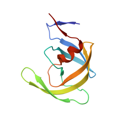
Fragment-based campaign for the identification of potential exosite binders of HIV-1 Protease

Forli, S.Tiefenbrunn, T.Baksh, M.M.Chang, M.W.Perryman, A.Garg, D.Happer, M.Lin, Y.-C.Goodsell, D.Angelina, E.L.De Vera, I.Kojetin, D.Torbett, B.E.Finn, M.G.Elder, J.Stout, C.D.Olson, A.

To be published.

Macromolecules
Find similar proteins by:  (by identity cutoff)  |  3D Structure
Entity ID: 1
MoleculeChains Sequence LengthOrganismDetailsImage
Protease
A, B
99Human immunodeficiency virus 1Mutation(s): 1 
Gene Names: pol
EC: 3.4.23.16
UniProt
Find proteins for P12497 (Human immunodeficiency virus type 1 group M subtype B (isolate NY5))
Explore P12497 
Go to UniProtKB:  P12497
Entity Groups  
Sequence Clusters30% Identity50% Identity70% Identity90% Identity95% Identity100% Identity
UniProt GroupP12497
Sequence Annotations
Expand
  • Reference Sequence
Small Molecules
Ligands 2 Unique
IDChains Name / Formula / InChI Key2D Diagram3D Interactions
3TL
Query on 3TL

Download Ideal Coordinates CCD File 
C [auth A]benzyl [(1S,4S,7S,8R,9R,10S,13S,16S)-7,10-dibenzyl-8,9-dihydroxy-1,16-dimethyl-4,13-bis(1-methylethyl)-2,5,12,15,18-pentaoxo-20-phenyl-19-oxa-3,6,11,14,17-pentaazaicos-1-yl]carbamate
C50 H64 N6 O10
BJJPNOGMLLUCER-KUTQPOQPSA-N
7GC
Query on 7GC

Download Ideal Coordinates CCD File 
D [auth A],
E [auth B]
4-methyl-N-(thiazol-2-ylcarbamoyl)benzenesulfonamide
C11 H11 N3 O3 S2
ZXPLKYLPCUVOFS-UHFFFAOYSA-N
Binding Affinity Annotations 
IDSourceBinding Affinity
3TL BindingDB:  5VCK Ki: 1.5 (nM) from 1 assay(s)
Biologically Interesting Molecules (External Reference) 1 Unique
Experimental Data & Validation

Experimental Data

  • Method: X-RAY DIFFRACTION
  • Resolution: 1.80 Å
  • R-Value Free: 0.214 
  • R-Value Work: 0.173 
  • R-Value Observed: 0.175 
  • Space Group: P 61
Unit Cell:
Length ( Å )Angle ( ˚ )
a = 62.953α = 90
b = 62.953β = 90
c = 82.306γ = 120
Software Package:
Software NamePurpose
SCALAdata scaling
REFMACrefinement
PDB_EXTRACTdata extraction
PHASERphasing
autoXDSdata reduction

Structure Validation

View Full Validation Report


Ligand Structure Quality Assessment 


Entry History & Funding Information

Deposition Data


Funding OrganizationLocationGrant Number
National Institutes of Health/National Institute of General Medical Sciences (NIH/NIGMS)United States1P50GM103368

Revision History  (Full details and data files)

  • Version 1.0: 2018-04-18
    Type: Initial release
  • Version 1.1: 2020-01-01
    Changes: Author supporting evidence
  • Version 1.2: 2023-10-04
    Changes: Data collection, Database references, Refinement description