5UE0

1.90 A resolution structure of CT622 C-terminal domain from Chlamydia trachomatis


Experimental Data Snapshot

  • Method: X-RAY DIFFRACTION
  • Resolution: 1.90 Å
  • R-Value Free: 0.258 
  • R-Value Work: 0.211 
  • R-Value Observed: 0.213 

wwPDB Validation   3D Report Full Report


This is version 1.4 of the entry. See complete history


Literature

The Loss of Expression of a Single Type 3 Effector (CT622) Strongly ReducesChlamydia trachomatisInfectivity and Growth.

Cosse, M.M.Barta, M.L.Fisher, D.J.Oesterlin, L.K.Niragire, B.Perrinet, S.Millot, G.A.Hefty, P.S.Subtil, A.

(2018) Front Cell Infect Microbiol 8: 145-145

  • DOI: https://doi.org/10.3389/fcimb.2018.00145
  • Primary Citation of Related Structures:  
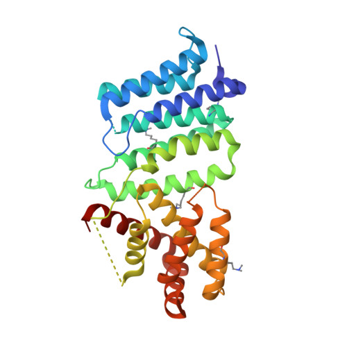
    5UE0

  • PubMed Abstract: 

    Invasion of epithelial cells by the obligate intracellular bacterium Chlamydia trachomatis results in its enclosure inside a membrane-bound compartment termed an inclusion. The bacterium quickly begins manipulating interactions between host intracellular trafficking and the inclusion interface, diverging from the endocytic pathway and escaping lysosomal fusion. We have identified a previously uncharacterized protein, CT622, unique to the Chlamydiaceae , in the absence of which most bacteria failed to establish a successful infection. CT622 is abundant in the infectious form of the bacteria, in which it associates with CT635, a putative novel chaperone protein. We show that CT622 is translocated into the host cytoplasm via type three secretion throughout the developmental cycle of the bacteria. Two separate domains of roughly equal size have been identified within CT622 and a 1.9 Å crystal structure of the C-terminal domain has been determined. Genetic disruption of ct622 expression resulted in a strong bacterial growth defect, which was due to deficiencies in proliferation and in the generation of infectious bacteria. Our results converge to identify CT622 as a secreted protein that plays multiple and crucial roles in the initiation and support of the C. trachomatis growth cycle. They reveal that genetic disruption of a single effector can deeply affect bacterial fitness.


  • Organizational Affiliation

    Unité de Biologie Cellulaire de l'Infection Microbienne, Institut Pasteur, Paris, France.


Macromolecules
Find similar proteins by:  (by identity cutoff)  |  3D Structure
Entity ID: 1
MoleculeChains Sequence LengthOrganismDetailsImage
CT622 protein299Chlamydia trachomatis L2/434/BuMutation(s): 0 
Gene Names: CTL0886
UniProt
Find proteins for A0A0H3MDV9 (Chlamydia trachomatis serovar L2 (strain 434/Bu / ATCC VR-902B))
Explore A0A0H3MDV9 
Go to UniProtKB:  A0A0H3MDV9
Entity Groups  
Sequence Clusters30% Identity50% Identity70% Identity90% Identity95% Identity100% Identity
UniProt GroupA0A0H3MDV9
Sequence Annotations
Expand
  • Reference Sequence
Small Molecules
Ligands 1 Unique
IDChains Name / Formula / InChI Key2D Diagram3D Interactions
SO4
Query on SO4

Download Ideal Coordinates CCD File 
B [auth A]SULFATE ION
O4 S
QAOWNCQODCNURD-UHFFFAOYSA-L
Modified Residues  1 Unique
IDChains TypeFormula2D DiagramParent
MLY
Query on MLY
A
L-PEPTIDE LINKINGC8 H18 N2 O2LYS
Experimental Data & Validation

Experimental Data

  • Method: X-RAY DIFFRACTION
  • Resolution: 1.90 Å
  • R-Value Free: 0.258 
  • R-Value Work: 0.211 
  • R-Value Observed: 0.213 
  • Space Group: I 2 3
Unit Cell:
Length ( Å )Angle ( ˚ )
a = 121.812α = 90
b = 121.812β = 90
c = 121.812γ = 90
Software Package:
Software NamePurpose
PHENIXrefinement
SCALAdata scaling
PHENIXphasing

Structure Validation

View Full Validation Report


Entry History & Funding Information

Deposition Data


Funding OrganizationLocationGrant Number
National Institutes of Health/National Institute Of Allergy and Infectious Diseases (NIH/NIAID)United StatesR01 AI079083

Revision History  (Full details and data files)

  • Version 1.0: 2018-01-10
    Type: Initial release
  • Version 1.1: 2018-05-30
    Changes: Data collection, Database references
  • Version 1.2: 2018-06-20
    Changes: Data collection, Database references
  • Version 1.3: 2018-07-25
    Changes: Data collection, Structure summary
  • Version 1.4: 2019-12-11
    Changes: Author supporting evidence