5U94

Crystal structure of the Mycobacterium tuberculosis PASTA kinase PknB in complex with the potential theraputic kinase inhibitor GSK690693.


Experimental Data Snapshot

  • Method: X-RAY DIFFRACTION
  • Resolution: 2.20 Å
  • R-Value Free: 0.237 
  • R-Value Work: 0.201 
  • R-Value Observed: 0.204 

wwPDB Validation   3D Report Full Report


Ligand Structure Quality Assessment 


This is version 1.5 of the entry. See complete history


Literature

In Silico Screen and Structural Analysis Identifies Bacterial Kinase Inhibitors which Act with beta-Lactams To Inhibit Mycobacterial Growth.

Wlodarchak, N.Teachout, N.Beczkiewicz, J.Procknow, R.Schaenzer, A.J.Satyshur, K.Pavelka, M.Zuercher, W.Drewry, D.Sauer, J.D.Striker, R.

(2018) Mol Pharm 15: 5410-5426

  • DOI: https://doi.org/10.1021/acs.molpharmaceut.8b00905
  • Primary Citation of Related Structures:  
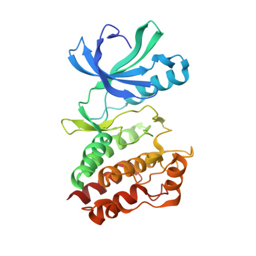
    5U94

  • PubMed Abstract: 

    New tools and concepts are needed to combat antimicrobial resistance. Actinomycetes and firmicutes share several eukaryotic-like Ser/Thr kinases (eSTK) that offer antibiotic development opportunities, including PknB, an essential mycobacterial eSTK. Despite successful development of potent biochemical PknB inhibitors by many groups, clinically useful microbiologic activity has been elusive. Additionally, PknB kinetics are not fully described, nor are structures with specific inhibitors available to inform inhibitor design. We used computational modeling with available structural information to identify human kinase inhibitors predicted to bind PknB, and we selected hits based on drug-like characteristics intended to increase the likelihood of cell entry. The computational model suggested a family of inhibitors, the imidazopyridine aminofurazans (IPAs), bind PknB with high affinity. We performed an in-depth characterization of PknB and found that these inhibitors biochemically inhibit PknB, with potency roughly following the predicted models. A novel X-ray structure confirmed that the inhibitors bound as predicted and made favorable protein contacts with the target. These inhibitors also have antimicrobial activity toward mycobacteria and nocardia. We demonstrated that the inhibitors are uniquely potentiated by β-lactams but not antibiotics traditionally used to treat mycobacteria, consistent with PknB's role in sensing cell wall stress. This is the first demonstration in the phylum actinobacteria that some β-lactam antibiotics could be more effective if paired with a PknB inhibitor. Collectively, our data show that in silico modeling can be used as a tool to discover promising drug leads, and the inhibitors we discovered can act with clinically relevant antibiotics to restore their efficacy against bacteria with limited treatment options.


  • Organizational Affiliation

    Department of Medicine , University of Wisconsin-Madison , 3341 Microbial Sciences Building, 1550 Linden Dr. , Madison , Wisconsin 53706 , United States.


Macromolecules
Find similar proteins by:  (by identity cutoff)  |  3D Structure
Entity ID: 1
MoleculeChains Sequence LengthOrganismDetailsImage
Serine/threonine-protein kinase PknB285Mycobacterium tuberculosis H37RvMutation(s): 0 
Gene Names: pknBRv0014cMTCY10H4.14c
EC: 2.7.11.1
UniProt
Find proteins for P9WI81 (Mycobacterium tuberculosis (strain ATCC 25618 / H37Rv))
Explore P9WI81 
Go to UniProtKB:  P9WI81
Entity Groups  
Sequence Clusters30% Identity50% Identity70% Identity90% Identity95% Identity100% Identity
UniProt GroupP9WI81
Sequence Annotations
Expand
  • Reference Sequence
Small Molecules
Ligands 3 Unique
IDChains Name / Formula / InChI Key2D Diagram3D Interactions
G93
Query on G93

Download Ideal Coordinates CCD File 
B [auth A]4-{2-(4-amino-1,2,5-oxadiazol-3-yl)-1-ethyl-7-[(3S)-piperidin-3-ylmethoxy]-1H-imidazo[4,5-c]pyridin-4-yl}-2-methylbut-3 -yn-2-ol
C21 H27 N7 O3
KGPGFQWBCSZGEL-ZDUSSCGKSA-N
GOL
Query on GOL

Download Ideal Coordinates CCD File 
C [auth A]
D [auth A]
E [auth A]
F [auth A]
G [auth A]
C [auth A],
D [auth A],
E [auth A],
F [auth A],
G [auth A],
H [auth A],
I [auth A],
J [auth A]
GLYCEROL
C3 H8 O3
PEDCQBHIVMGVHV-UHFFFAOYSA-N
MG
Query on MG

Download Ideal Coordinates CCD File 
K [auth A]MAGNESIUM ION
Mg
JLVVSXFLKOJNIY-UHFFFAOYSA-N
Binding Affinity Annotations 
IDSourceBinding Affinity
G93 BindingDB:  5U94 Kd: 3.2 (nM) from 1 assay(s)
Experimental Data & Validation

Experimental Data

  • Method: X-RAY DIFFRACTION
  • Resolution: 2.20 Å
  • R-Value Free: 0.237 
  • R-Value Work: 0.201 
  • R-Value Observed: 0.204 
  • Space Group: C 2 2 21
Unit Cell:
Length ( Å )Angle ( ˚ )
a = 114.786α = 90
b = 121.294β = 90
c = 49.529γ = 90
Software Package:
Software NamePurpose
SCALEPACKdata scaling
PHENIXrefinement
PDB_EXTRACTdata extraction
HKL-2000data reduction
PHENIXphasing

Structure Validation

View Full Validation Report


Ligand Structure Quality Assessment 


Entry History & Funding Information

Deposition Data


Funding OrganizationLocationGrant Number
National Institutes of Health/National Center for Advancing Translational Sciences (NIH/NCATS)United StatesUL1TR000427
Hartwell FoundationUnited States--

Revision History  (Full details and data files)

  • Version 1.0: 2017-12-20
    Type: Initial release
  • Version 1.1: 2018-01-17
    Changes: Author supporting evidence
  • Version 1.2: 2018-04-18
    Changes: Data collection
  • Version 1.3: 2019-01-16
    Changes: Data collection, Database references
  • Version 1.4: 2019-12-04
    Changes: Author supporting evidence
  • Version 1.5: 2023-10-04
    Changes: Data collection, Database references, Derived calculations, Refinement description, Structure summary