5U56

Structure of Francisella tularensis heterodimeric SspA (MglA-SspA)


Experimental Data Snapshot

  • Method: X-RAY DIFFRACTION
  • Resolution: 2.65 Å
  • R-Value Free: 0.259 
  • R-Value Work: 0.204 
  • R-Value Observed: 0.207 

wwPDB Validation   3D Report Full Report


This is version 1.2 of the entry. See complete history


Literature

Dissection of the molecular circuitry controlling virulence in Francisella tularensis.

Cuthbert, B.J.Ross, W.Rohlfing, A.E.Dove, S.L.Gourse, R.L.Brennan, R.G.Schumacher, M.A.

(2017) Genes Dev 31: 1549-1560

  • DOI: https://doi.org/10.1101/gad.303701.117
  • Primary Citation of Related Structures:  
    5U51, 5U56, 6ALX

  • PubMed Abstract: 

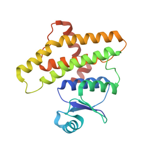
    Francisella tularensis, the etiological agent of tularemia, is one of the most infectious bacteria known. Because of its extreme pathogenicity, F. tularensis is classified as a category A bioweapon by the US government. F. tularensis virulence stems from genes encoded on the Francisella pathogenicity island (FPI). An unusual set of Francisella regulators-the heteromeric macrophage growth locus protein A (MglA)-stringent starvation protein A (SspA) complex and the DNA-binding protein pathogenicity island gene regulator (PigR)-activates FPI transcription and thus is essential for virulence. Intriguingly, the second messenger, guanosine-tetraphosphate (ppGpp), which is produced during infection, is also involved in coordinating Francisella virulence; however, its role has been unclear. Here we identify MglA-SspA as a novel ppGpp-binding complex and describe structures of apo- and ppGpp-bound MglA-SspA. We demonstrate that MglA-SspA, which binds RNA polymerase (RNAP), also interacts with the C-terminal domain of PigR, thus anchoring the (MglA-SspA)-RNAP complex to the FPI promoter. Furthermore, we show that MglA-SspA must be bound to ppGpp to mediate high-affinity interactions with PigR. Thus, these studies unveil a novel pathway different from those described previously for regulation of transcription by ppGpp. The data also indicate that F. tularensis pathogenesis is controlled by a highly interconnected molecular circuitry in which the virulence machinery directly senses infection via a small molecule stress signal.


  • Organizational Affiliation

    Department of Biochemistry, Duke University School of Medicine, Durham, North Carolina 27710, USA.


Macromolecules
Find similar proteins by:  (by identity cutoff)  |  3D Structure
Entity ID: 1
MoleculeChains Sequence LengthOrganismDetailsImage
Stringent starvation protein AA [auth C],
C [auth D]
211Francisella tularensisMutation(s): 0 
Gene Names: DR86_1150
UniProt
Find proteins for Q5NHJ6 (Francisella tularensis subsp. tularensis (strain SCHU S4 / Schu 4))
Explore Q5NHJ6 
Go to UniProtKB:  Q5NHJ6
Entity Groups  
Sequence Clusters30% Identity50% Identity70% Identity90% Identity95% Identity100% Identity
UniProt GroupQ5NHJ6
Sequence Annotations
Expand
  • Reference Sequence
Find similar proteins by:  (by identity cutoff)  |  3D Structure
Entity ID: 2
MoleculeChains Sequence LengthOrganismDetailsImage
Macrophage growth locus AB [auth A],
D [auth B]
204Francisella tularensisMutation(s): 0 
Gene Names: DR86_1530
UniProt
Find proteins for Q5NFG1 (Francisella tularensis subsp. tularensis (strain SCHU S4 / Schu 4))
Explore Q5NFG1 
Go to UniProtKB:  Q5NFG1
Entity Groups  
Sequence Clusters30% Identity50% Identity70% Identity90% Identity95% Identity100% Identity
UniProt GroupQ5NFG1
Sequence Annotations
Expand
  • Reference Sequence
Small Molecules
Ligands 3 Unique
IDChains Name / Formula / InChI Key2D Diagram3D Interactions
1PE
Query on 1PE

Download Ideal Coordinates CCD File 
G [auth C],
J [auth A]
PENTAETHYLENE GLYCOL
C10 H22 O6
JLFNLZLINWHATN-UHFFFAOYSA-N
PRO
Query on PRO

Download Ideal Coordinates CCD File 
K [auth A],
Q [auth B]
PROLINE
C5 H9 N O2
ONIBWKKTOPOVIA-BYPYZUCNSA-N
GOL
Query on GOL

Download Ideal Coordinates CCD File 
E [auth C]
F [auth C]
H [auth A]
I [auth A]
L [auth D]
E [auth C],
F [auth C],
H [auth A],
I [auth A],
L [auth D],
M [auth D],
N [auth D],
O [auth B],
P [auth B]
GLYCEROL
C3 H8 O3
PEDCQBHIVMGVHV-UHFFFAOYSA-N
Experimental Data & Validation

Experimental Data

  • Method: X-RAY DIFFRACTION
  • Resolution: 2.65 Å
  • R-Value Free: 0.259 
  • R-Value Work: 0.204 
  • R-Value Observed: 0.207 
  • Space Group: P 21 21 21
Unit Cell:
Length ( Å )Angle ( ˚ )
a = 66.408α = 90
b = 113.261β = 90
c = 140.4γ = 90
Software Package:
Software NamePurpose
PHENIXrefinement
HKL-2000data reduction
HKL-2000data scaling
PHASERphasing

Structure Validation

View Full Validation Report


Entry History 

Deposition Data

Revision History  (Full details and data files)

  • Version 1.0: 2017-08-16
    Type: Initial release
  • Version 1.1: 2017-09-27
    Changes: Database references
  • Version 1.2: 2023-10-04
    Changes: Data collection, Database references, Refinement description