5TGV

Crystal structure of H10 hemagglutinin mutant (K158aA-D193T-Q226L-G228S) from Jiangxi-Donghu (2013) H10N8 influenza virus in complex with 3'-SLN


Experimental Data Snapshot

  • Method: X-RAY DIFFRACTION
  • Resolution: 2.97 Å
  • R-Value Free: 0.260 
  • R-Value Work: 0.187 
  • R-Value Observed: 0.191 

wwPDB Validation   3D Report Full Report


Ligand Structure Quality Assessment 


This is version 2.1 of the entry. See complete history


Literature

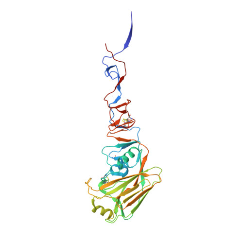
The 150-Loop Restricts the Host Specificity of Human H10N8 Influenza Virus.

Tzarum, N.de Vries, R.P.Peng, W.Thompson, A.J.Bouwman, K.M.McBride, R.Yu, W.Zhu, X.Verheije, M.H.Paulson, J.C.Wilson, I.A.

(2017) Cell Rep 19: 235-245

  • DOI: https://doi.org/10.1016/j.celrep.2017.03.054
  • Primary Citation of Related Structures:  
    5TGO, 5TGU, 5TGV, 5TH0, 5TH1, 5THB, 5THC, 5THF

  • PubMed Abstract: 

    Adaptation of influenza A viruses to new hosts are rare events but are the basis for emergence of new influenza pandemics in the human population. Thus, understanding the processes involved in such events is critical for anticipating potential pandemic threats. In 2013, the first case of human infection by an avian H10N8 virus was reported, yet the H10 hemagglutinin (HA) maintains avian receptor specificity. However, the 150-loop of H10 HA, as well as related H7 and H15 subtypes, contains a two-residue insert that can potentially block human receptor binding. Mutation of the 150-loop on the background of Q226L and G228S mutations, which arose in the receptor-binding site of human pandemic H2 and H3 viruses, resulted in acquisition of human-type receptor specificity. Crystal structures of H10 HA mutants with human and avian receptor analogs, receptor-binding studies, and tissue staining experiments illustrate the important role of the 150-loop in H10 receptor specificity.


  • Organizational Affiliation

    Department of Integrative Structural and Computational Biology, The Scripps Research Institute, 10550 North Torrey Pines Road, La Jolla, CA 92037, USA.


Macromolecules
Find similar proteins by:  (by identity cutoff)  |  3D Structure
Entity ID: 1
MoleculeChains Sequence LengthOrganismDetailsImage
Hemagglutinin HA1 chain
A, C, E
323Influenza A virusMutation(s): 4 
UniProt
Find proteins for A0A059T4A1 (Influenza A virus)
Explore A0A059T4A1 
Go to UniProtKB:  A0A059T4A1
Entity Groups  
Sequence Clusters30% Identity50% Identity70% Identity90% Identity95% Identity100% Identity
UniProt GroupA0A059T4A1
Sequence Annotations
Expand
  • Reference Sequence
Find similar proteins by:  (by identity cutoff)  |  3D Structure
Entity ID: 2
MoleculeChains Sequence LengthOrganismDetailsImage
Hemagglutinin HA2 chain
B, D, F
180Influenza A virusMutation(s): 0 
UniProt
Find proteins for A0A059T4A1 (Influenza A virus)
Explore A0A059T4A1 
Go to UniProtKB:  A0A059T4A1
Entity Groups  
Sequence Clusters30% Identity50% Identity70% Identity90% Identity95% Identity100% Identity
UniProt GroupA0A059T4A1
Sequence Annotations
Expand
  • Reference Sequence
Oligosaccharides

Help

Entity ID: 3
MoleculeChains Length2D Diagram Glycosylation3D Interactions
2-acetamido-2-deoxy-beta-D-glucopyranose-(1-4)-2-acetamido-2-deoxy-beta-D-glucopyranose
G
2N-Glycosylation
Glycosylation Resources
GlyTouCan:  G42666HT
GlyCosmos:  G42666HT
GlyGen:  G42666HT
Entity ID: 4
MoleculeChains Length2D Diagram Glycosylation3D Interactions
N-acetyl-alpha-neuraminic acid-(2-3)-beta-D-galactopyranose-(1-4)-2-acetamido-2-deoxy-beta-D-glucopyranose
H
3N/A
Glycosylation Resources
GlyTouCan:  G00065MO
GlyCosmos:  G00065MO
GlyGen:  G00065MO
Entity ID: 5
MoleculeChains Length2D Diagram Glycosylation3D Interactions
beta-D-mannopyranose-(1-4)-2-acetamido-2-deoxy-beta-D-glucopyranose
I
2N/A
Glycosylation Resources
GlyTouCan:  G90333CG
GlyCosmos:  G90333CG
GlyGen:  G90333CG
Biologically Interesting Molecules (External Reference) 1 Unique
Experimental Data & Validation

Experimental Data

  • Method: X-RAY DIFFRACTION
  • Resolution: 2.97 Å
  • R-Value Free: 0.260 
  • R-Value Work: 0.187 
  • R-Value Observed: 0.191 
  • Space Group: P 1 21 1
Unit Cell:
Length ( Å )Angle ( ˚ )
a = 63.512α = 90
b = 253.173β = 111.97
c = 69.964γ = 90
Software Package:
Software NamePurpose
PHENIXrefinement
HKL-2000data reduction
HKL-2000data scaling
PHASERphasing

Structure Validation

View Full Validation Report


Ligand Structure Quality Assessment 


Entry History 

Deposition Data

Revision History  (Full details and data files)

  • Version 1.0: 2017-04-05
    Type: Initial release
  • Version 1.1: 2017-04-26
    Changes: Database references
  • Version 1.2: 2017-11-22
    Changes: Refinement description
  • Version 2.0: 2020-07-29
    Type: Remediation
    Reason: Carbohydrate remediation
    Changes: Advisory, Atomic model, Data collection, Derived calculations, Structure summary
  • Version 2.1: 2023-10-04
    Changes: Data collection, Database references, Derived calculations, Refinement description, Structure summary