5SYB

Crystal structure of human PHF5A


Experimental Data Snapshot

  • Method: X-RAY DIFFRACTION
  • Resolution: 1.82 Å
  • R-Value Free: 0.203 
  • R-Value Work: 0.168 
  • R-Value Observed: 0.170 

wwPDB Validation   3D Report Full Report


This is version 1.3 of the entry. See complete history


Literature

Splicing modulators act at the branch point adenosine binding pocket defined by the PHF5A-SF3b complex.

Teng, T.Tsai, J.H.Puyang, X.Seiler, M.Peng, S.Prajapati, S.Aird, D.Buonamici, S.Caleb, B.Chan, B.Corson, L.Feala, J.Fekkes, P.Gerard, B.Karr, C.Korpal, M.Liu, X.T Lowe, J.Mizui, Y.Palacino, J.Park, E.Smith, P.G.Subramanian, V.Wu, Z.J.Zou, J.Yu, L.Chicas, A.Warmuth, M.Larsen, N.Zhu, P.

(2017) Nat Commun 8: 15522-15522

  • DOI: https://doi.org/10.1038/ncomms15522
  • Primary Citation of Related Structures:  
    5SYB

  • PubMed Abstract: 

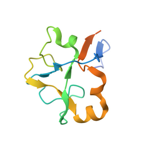
    Pladienolide, herboxidiene and spliceostatin have been identified as splicing modulators that target SF3B1 in the SF3b subcomplex. Here we report that PHF5A, another component of this subcomplex, is also targeted by these compounds. Mutations in PHF5A-Y36, SF3B1-K1071, SF3B1-R1074 and SF3B1-V1078 confer resistance to these modulators, suggesting a common interaction site. RNA-seq analysis reveals that PHF5A-Y36C has minimal effect on basal splicing but inhibits the global action of splicing modulators. Moreover, PHF5A-Y36C alters splicing modulator-induced intron-retention/exon-skipping profile, which correlates with the differential GC content between adjacent introns and exons. We determine the crystal structure of human PHF5A demonstrating that Y36 is located on a highly conserved surface. Analysis of the cryo-EM spliceosome B act complex shows that the resistance mutations cluster in a pocket surrounding the branch point adenosine, suggesting a competitive mode of action. Collectively, we propose that PHF5A-SF3B1 forms a central node for binding to these splicing modulators.


  • Organizational Affiliation

    H3 Biomedicine Inc., 300 Technology Sq, 5th Floor, Cambridge, Massachusetts 02139, USA.


Macromolecules
Find similar proteins by:  (by identity cutoff)  |  3D Structure
Entity ID: 1
MoleculeChains Sequence LengthOrganismDetailsImage
PHD finger-like domain-containing protein 5A
A, B
113Homo sapiensMutation(s): 1 
Gene Names: PHF5A
UniProt & NIH Common Fund Data Resources
Find proteins for Q7RTV0 (Homo sapiens)
Explore Q7RTV0 
Go to UniProtKB:  Q7RTV0
PHAROS:  Q7RTV0
GTEx:  ENSG00000100410 
Entity Groups  
Sequence Clusters30% Identity50% Identity70% Identity90% Identity95% Identity100% Identity
UniProt GroupQ7RTV0
Sequence Annotations
Expand
  • Reference Sequence
Small Molecules
Ligands 2 Unique
IDChains Name / Formula / InChI Key2D Diagram3D Interactions
ZN
Query on ZN

Download Ideal Coordinates CCD File 
C [auth A]
D [auth A]
E [auth A]
H [auth B]
I [auth B]
C [auth A],
D [auth A],
E [auth A],
H [auth B],
I [auth B],
J [auth B]
ZINC ION
Zn
PTFCDOFLOPIGGS-UHFFFAOYSA-N
EDO
Query on EDO

Download Ideal Coordinates CCD File 
F [auth A],
G [auth A],
K [auth B],
L [auth B]
1,2-ETHANEDIOL
C2 H6 O2
LYCAIKOWRPUZTN-UHFFFAOYSA-N
Experimental Data & Validation

Experimental Data

  • Method: X-RAY DIFFRACTION
  • Resolution: 1.82 Å
  • R-Value Free: 0.203 
  • R-Value Work: 0.168 
  • R-Value Observed: 0.170 
  • Space Group: P 21 3
Unit Cell:
Length ( Å )Angle ( ˚ )
a = 82.116α = 90
b = 82.116β = 90
c = 82.116γ = 90
Software Package:
Software NamePurpose
REFMACrefinement
xia2data reduction
xia2data scaling
SHELXDEphasing

Structure Validation

View Full Validation Report


Entry History 

Deposition Data

Revision History  (Full details and data files)

  • Version 1.0: 2016-09-07
    Type: Initial release
  • Version 1.1: 2016-10-26
    Changes: Structure summary
  • Version 1.2: 2017-06-07
    Changes: Database references
  • Version 1.3: 2024-03-06
    Changes: Data collection, Database references