5SIC

MOLECULAR RECOGNITION AT THE ACTIVE SITE OF SUBTILISIN BPN': CRYSTALLOGRAPHIC STUDIES USING GENETICALLY ENGINEERED PROTEINACEOUS INHIBITOR SSI (STREPTOMYCES SUBTILISIN INHIBITOR)


Experimental Data Snapshot

  • Method: X-RAY DIFFRACTION
  • Resolution: 2.20 Å
  • R-Value Observed: 0.176 

wwPDB Validation   3D Report Full Report


This is version 1.3 of the entry. See complete history


Literature

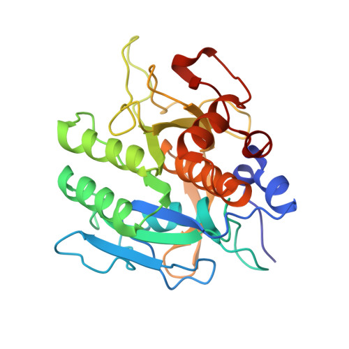
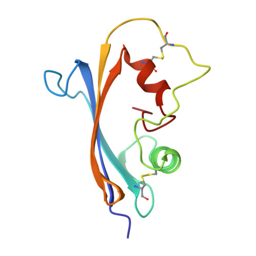
Molecular recognition at the active site of subtilisin BPN': crystallographic studies using genetically engineered proteinaceous inhibitor SSI (Streptomyces subtilisin inhibitor).

Takeuchi, Y.Noguchi, S.Satow, Y.Kojima, S.Kumagai, I.Miura, K.Nakamura, K.T.Mitsui, Y.

(1991) Protein Eng 4: 501-508

  • DOI: https://doi.org/10.1093/protein/4.5.501
  • Primary Citation of Related Structures:  
    3SIC, 5SIC

  • PubMed Abstract: 

    Unlike trypsin-like serine proteases having only one conspicuous binding pocket in the active site, subtilisin BPN' has two such pockets, the S1 and S4 pockets, which accommodate the P1 and P4 residues of ligands (after Schechter and Berger notation) respectively. Using computer graphics, the geometrical nature of the two pockets was carefully examined and strategies for site-directed mutagenesis studies were set up against a protein SSI (Streptomyces subtilisin inhibitor), which is a strong proteinaceous inhibitor (or a substrate analogue) of subtilisin BPN'. It was decided to convert the P1 residue, methionine 73, into lysine (M73K) with or without additional conversion of the P4 residue, methionine 70, into glycine (M70G). The crystal structures of the two complexes of subtilisin BPN', one with the single mutant SSI (M73K) and the other with the double mutant SSI (M73K, M70G) were solved showing that (i) small 'electrostatic induced-fit movement' occurs in the S1 pocket upon introducing the terminal plus charge of the lysine side chain, and (ii) large 'mechanical induced-fit movement' occurs in the S4 pocket upon reducing the size of the P4 side chain from methionine to glycine. In both (i) and (ii), the induced-fit movement occurred in a concerted fashion involving both the enzyme and 'substrate' amino acid residues. The term 'substrate-assisted stabilization' was coined to stress the cooperative nature of the induced-fit movements.


  • Organizational Affiliation

    Pharmaceutical Research Center, Meiji Seika Kaisha, Ltd., Yokohama, Japan.


Macromolecules
Find similar proteins by:  (by identity cutoff)  |  3D Structure
Entity ID: 1
MoleculeChains Sequence LengthOrganismDetailsImage
SUBTILISIN BPN'A [auth E]275Bacillus amyloliquefaciensMutation(s): 0 
EC: 3.4.21.62
UniProt
Find proteins for P00782 (Bacillus amyloliquefaciens)
Explore P00782 
Go to UniProtKB:  P00782
Entity Groups  
Sequence Clusters30% Identity50% Identity70% Identity90% Identity95% Identity100% Identity
UniProt GroupP00782
Sequence Annotations
Expand
  • Reference Sequence
Find similar proteins by:  (by identity cutoff)  |  3D Structure
Entity ID: 2
MoleculeChains Sequence LengthOrganismDetailsImage
SUBTILISIN INHIBITOR (SSI)B [auth I]107Streptomyces albogriseolusMutation(s): 0 
Gene Names: ssi
UniProt
Find proteins for P01006 (Streptomyces albogriseolus)
Explore P01006 
Go to UniProtKB:  P01006
Entity Groups  
Sequence Clusters30% Identity50% Identity70% Identity90% Identity95% Identity100% Identity
UniProt GroupP01006
Sequence Annotations
Expand
  • Reference Sequence
Small Molecules
Ligands 1 Unique
IDChains Name / Formula / InChI Key2D Diagram3D Interactions
CA
Query on CA

Download Ideal Coordinates CCD File 
C [auth E],
D [auth E]
CALCIUM ION
Ca
BHPQYMZQTOCNFJ-UHFFFAOYSA-N
Experimental Data & Validation

Experimental Data

  • Method: X-RAY DIFFRACTION
  • Resolution: 2.20 Å
  • R-Value Observed: 0.176 
  • Space Group: I 2 2 2
Unit Cell:
Length ( Å )Angle ( ˚ )
a = 77.2α = 90
b = 185.9β = 90
c = 69.5γ = 90
Software Package:
Software NamePurpose
PROLSQrefinement

Structure Validation

View Full Validation Report


Entry History 

Deposition Data

Revision History  (Full details and data files)

  • Version 1.0: 1994-01-31
    Type: Initial release
  • Version 1.1: 2008-03-03
    Changes: Version format compliance
  • Version 1.2: 2011-07-13
    Changes: Version format compliance
  • Version 1.3: 2017-11-29
    Changes: Derived calculations, Other