5OXF

An oligomerised bacterial dynamin pair provides a mechanism for the long range sensing and tethering of membranes


Experimental Data Snapshot

  • Method: X-RAY DIFFRACTION
  • Resolution: 3.94 Å
  • R-Value Free: 0.288 
  • R-Value Work: 0.259 
  • R-Value Observed: 0.260 

wwPDB Validation   3D Report Full Report


Ligand Structure Quality Assessment 


This is version 1.0 of the entry. See complete history


Literature

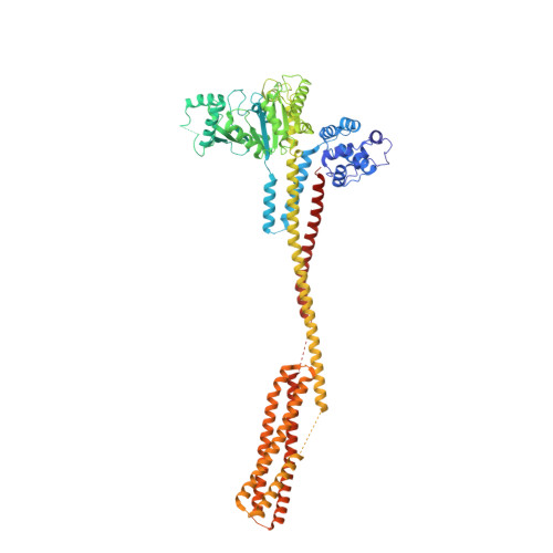
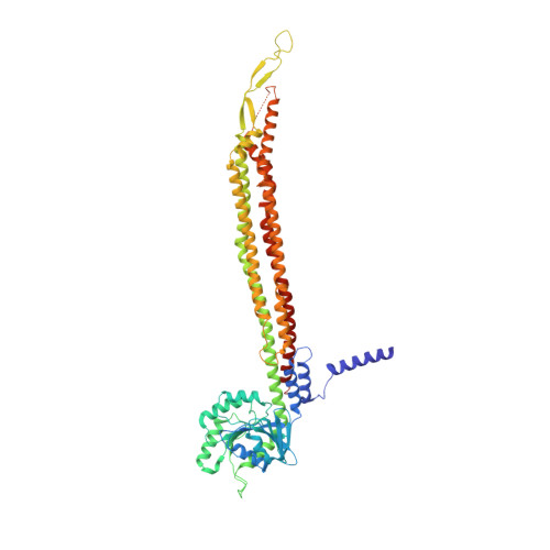
Structural basis for membrane tethering by a bacterial dynamin-like pair.

Liu, J.Noel, J.K.Low, H.H.

(2018) Nat Commun 9: 3345-3345

  • DOI: https://doi.org/10.1038/s41467-018-05523-8
  • Primary Citation of Related Structures:  
    5OWV, 5OXF

  • PubMed Abstract: 

    Dynamin-like proteins (DLPs) are large GTPases that restructure membrane. DLPs such as the mitofusins form heterotypic oligomers between isoform pairs that bridge and fuse opposing membranes. In bacteria, heterotypic oligomerisation may also be important for membrane remodelling as most DLP genes are paired within operons. How DLPs tether opposing membranes is unknown. Here we show the crystal structure of a DLP heterotypic pair from the pathogen Campylobacter jejuni. A 2:2 stoichiometric tetramer is observed where heterodimers, conjoined by a random coil linker, assemble back-to-back to form a tripartite DLP chain with extreme flexibility. In vitro, tetramerisation triggers GTPase activity and induces lipid binding. Liposomes are readily tethered and form tubes at high tetramer concentration. Our results provide a direct mechanism for the long-range binding and bridging of opposing membranes by a bacterial DLP pair. They also provide broad mechanistic and structural insights that are relevant to other heterotypic DLP complexes.


  • Organizational Affiliation

    Department of Life Sciences, Imperial College, London, SW7 2AZ, UK.


Macromolecules
Find similar proteins by:  (by identity cutoff)  |  3D Structure
Entity ID: 1
MoleculeChains Sequence LengthOrganismDetailsImage
GTP-binding protein
A, B
732Campylobacter jejuniMutation(s): 0 
Gene Names: BKM79_02020CRM98_01430
UniProt
Find proteins for A0A0H3PJL7 (Campylobacter jejuni subsp. jejuni serotype O:23/36 (strain 81-176))
Explore A0A0H3PJL7 
Go to UniProtKB:  A0A0H3PJL7
Entity Groups  
Sequence Clusters30% Identity50% Identity70% Identity90% Identity95% Identity100% Identity
UniProt GroupA0A0H3PJL7
Sequence Annotations
Expand
  • Reference Sequence
Find similar proteins by:  (by identity cutoff)  |  3D Structure
Entity ID: 2
MoleculeChains Sequence LengthOrganismDetailsImage
GTP-binding protein
C, D
614Campylobacter jejuniMutation(s): 0 
Gene Names: BKM79_02025
UniProt
Find proteins for A0A0H3PJK4 (Campylobacter jejuni subsp. jejuni serotype O:23/36 (strain 81-176))
Explore A0A0H3PJK4 
Go to UniProtKB:  A0A0H3PJK4
Entity Groups  
Sequence Clusters30% Identity50% Identity70% Identity90% Identity95% Identity100% Identity
UniProt GroupA0A0H3PJK4
Sequence Annotations
Expand
  • Reference Sequence
Experimental Data & Validation

Experimental Data

  • Method: X-RAY DIFFRACTION
  • Resolution: 3.94 Å
  • R-Value Free: 0.288 
  • R-Value Work: 0.259 
  • R-Value Observed: 0.260 
  • Space Group: P 21 21 21
Unit Cell:
Length ( Å )Angle ( ˚ )
a = 114.717α = 90
b = 228.675β = 90
c = 318.8γ = 90
Software Package:
Software NamePurpose
PHENIXrefinement
XDSdata reduction
Aimlessdata scaling
PHENIXphasing

Structure Validation

View Full Validation Report


Ligand Structure Quality Assessment 


Entry History & Funding Information

Deposition Data


Funding OrganizationLocationGrant Number
Wellcome TrustUnited Kingdom097328/Z/11/Z

Revision History  (Full details and data files)

  • Version 1.0: 2018-09-05
    Type: Initial release