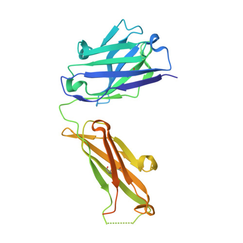
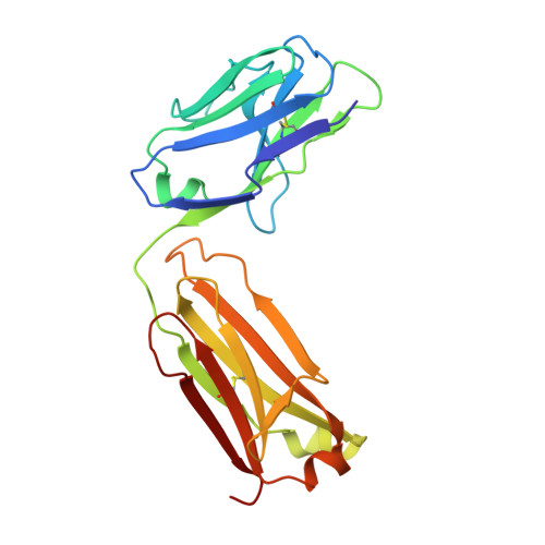
5OPY

Crystal structure of anti-alphaVbeta3 integrin Fab LM609


Experimental Data Snapshot

  • Method: X-RAY DIFFRACTION
  • Resolution: 2.26 Å
  • R-Value Free: 0.255 
  • R-Value Work: 0.213 
  • R-Value Observed: 0.215 

wwPDB Validation   3D Report Full Report


This is version 1.3 of the entry. See complete history


Literature

The Therapeutic Antibody LM609 Selectively Inhibits Ligand Binding to Human alpha V beta 3 Integrin via Steric Hindrance.

Borst, A.J.James, Z.M.Zagotta, W.N.Ginsberg, M.Rey, F.A.DiMaio, F.Backovic, M.Veesler, D.

(2017) Structure 25: 1732-1739.e5

  • DOI: https://doi.org/10.1016/j.str.2017.09.007
  • Primary Citation of Related Structures:  
    5OPY, 6AVQ, 6AVR, 6AVU

  • PubMed Abstract: 

    The LM609 antibody specifically recognizes α V β 3 integrin and inhibits angiogenesis, bone resorption, and viral infections in an arginine-glycine-aspartate-independent manner. LM609 entered phase II clinical trials for the treatment of several cancers and was also used for α V β 3 -targeted radioimmunotherapy. To elucidate the mechanisms of recognition and inhibition of α V β 3 integrin, we solved the structure of the LM609 antigen-binding fragment by X-ray crystallography and determined its binding affinity for α V β 3 . Using single-particle electron microscopy, we show that LM609 binds at the interface between the β-propeller domain of the α V chain and the βI domain of the β 3 chain, near the RGD-binding site, of all observed integrin conformational states. Integrating these data with fluorescence size-exclusion chromatography, we demonstrate that LM609 sterically hinders access of large ligands to the RGD-binding pocket, without obstructing it. This work provides a structural framework to expedite future efforts utilizing LM609 as a diagnostic or therapeutic tool.


  • Organizational Affiliation

    Department of Biochemistry, University of Washington, Seattle, WA 98195, USA.


Macromolecules
Find similar proteins by:  (by identity cutoff)  |  3D Structure
Entity ID: 1
MoleculeChains Sequence LengthOrganismDetailsImage
Heavy chain of LM609 Fab (antigen-binding fragment)A [auth H]257Mus musculusMutation(s): 0 
Entity Groups  
Sequence Clusters30% Identity50% Identity70% Identity90% Identity95% Identity100% Identity
Sequence Annotations
Expand
  • Reference Sequence
Find similar proteins by:  (by identity cutoff)  |  3D Structure
Entity ID: 2
MoleculeChains Sequence LengthOrganismDetailsImage
Light chain of LM609 Fab (antigen-binding fragment)B [auth L]214Mus musculusMutation(s): 0 
Entity Groups  
Sequence Clusters30% Identity50% Identity70% Identity90% Identity95% Identity100% Identity
Sequence Annotations
Expand
  • Reference Sequence
Experimental Data & Validation

Experimental Data

  • Method: X-RAY DIFFRACTION
  • Resolution: 2.26 Å
  • R-Value Free: 0.255 
  • R-Value Work: 0.213 
  • R-Value Observed: 0.215 
  • Space Group: P 2 21 21
Unit Cell:
Length ( Å )Angle ( ˚ )
a = 51.81α = 90
b = 82.35β = 90
c = 92.17γ = 90
Software Package:
Software NamePurpose
BUSTERrefinement
XDSdata reduction
Aimlessdata scaling
PHASERphasing

Structure Validation

View Full Validation Report


Entry History & Funding Information

Deposition Data


Funding OrganizationLocationGrant Number
Institut PasteurFranceRecurrent funding
National Institutes of Health/National Institute of General Medical Sciences (NIH/NIGMS)United States1R01GM120553
National Institutes of Health/National Institute of General Medical Sciences (NIH/NIGMS)United StatesT32GM008268

Revision History  (Full details and data files)

  • Version 1.0: 2017-10-25
    Type: Initial release
  • Version 1.1: 2017-11-15
    Changes: Database references
  • Version 1.2: 2022-03-30
    Changes: Author supporting evidence, Database references
  • Version 1.3: 2024-01-17
    Changes: Data collection, Refinement description