5OCW

Structure of Mycobacterium tuberculosis tryptophan synthase in space group F222


Experimental Data Snapshot

  • Method: X-RAY DIFFRACTION
  • Resolution: 4.00 Å
  • R-Value Free: 0.368 
  • R-Value Work: 0.358 
  • R-Value Observed: 0.358 

wwPDB Validation   3D Report Full Report


Ligand Structure Quality Assessment 


This is version 1.4 of the entry. See complete history


Literature

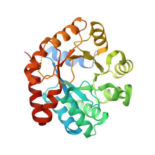
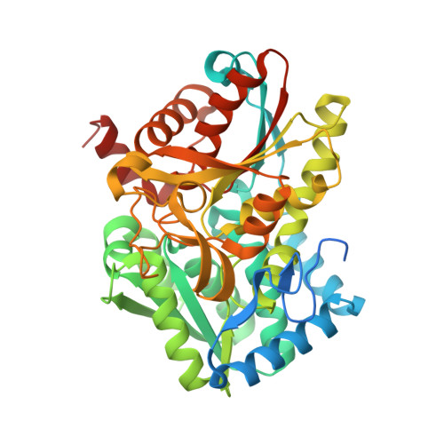
Inhibiting mycobacterial tryptophan synthase by targeting the inter-subunit interface.

Abrahams, K.A.Cox, J.A.G.Futterer, K.Rullas, J.Ortega-Muro, F.Loman, N.J.Moynihan, P.J.Perez-Herran, E.Jimenez, E.Esquivias, J.Barros, D.Ballell, L.Alemparte, C.Besra, G.S.

(2017) Sci Rep 7: 9430-9430

  • DOI: https://doi.org/10.1038/s41598-017-09642-y
  • Primary Citation of Related Structures:  
    5OCW

  • PubMed Abstract: 

    Drug discovery efforts against the pathogen Mycobacterium tuberculosis (Mtb) have been advanced through phenotypic screens of extensive compound libraries. Such a screen revealed sulfolane 1 and indoline-5-sulfonamides 2 and 3 as potent inhibitors of mycobacterial growth. Optimization in the sulfolane series led to compound 4, which has proven activity in an in vivo murine model of Mtb infection. Here we identify the target and mode of inhibition of these compounds based on whole genome sequencing of spontaneous resistant mutants, which identified mutations locating to the essential α- and β-subunits of tryptophan synthase. Over-expression studies confirmed tryptophan synthase as the biological target. Biochemical techniques probed the mechanism of inhibition, revealing the mutant enzyme complex incurs a fitness cost but does not prevent inhibitor binding. Mapping of the resistance conferring mutations onto a low-resolution crystal structure of Mtb tryptophan synthase showed they locate to the interface between the α- and β-subunits. The discovery of anti-tubercular agents inhibiting tryptophan synthase highlights the therapeutic potential of this enzyme and draws attention to the prospect of other amino acid biosynthetic pathways as future Mtb drug targets.


  • Organizational Affiliation

    Institute of Microbiology and Infection, School of Biosciences, University of Birmingham, Edgbaston, Birmingham, B15 2TT, UK.


Macromolecules
Find similar proteins by:  (by identity cutoff)  |  3D Structure
Entity ID: 1
MoleculeChains Sequence LengthOrganismDetailsImage
Tryptophan synthase alpha chain
A, C, E, G, I
A, C, E, G, I, K, M, O, Q, S, U, W
290Mycobacterium tuberculosis H37RvMutation(s): 0 
Gene Names: trpARv1613MTCY01B2.05
EC: 4.2.1.20
UniProt
Find proteins for P9WFY1 (Mycobacterium tuberculosis (strain ATCC 25618 / H37Rv))
Explore P9WFY1 
Go to UniProtKB:  P9WFY1
Entity Groups  
Sequence Clusters30% Identity50% Identity70% Identity90% Identity95% Identity100% Identity
UniProt GroupP9WFY1
Sequence Annotations
Expand
  • Reference Sequence
Find similar proteins by:  (by identity cutoff)  |  3D Structure
Entity ID: 2
MoleculeChains Sequence LengthOrganismDetailsImage
Tryptophan synthase beta chain
B, D, F, H, J
B, D, F, H, J, L, N, P, R, T, V, X
442Mycobacterium tuberculosis H37RvMutation(s): 0 
Gene Names: trpBRv1612MTCY01B2.04
EC: 4.2.1.20
UniProt
Find proteins for P9WFX9 (Mycobacterium tuberculosis (strain ATCC 25618 / H37Rv))
Explore P9WFX9 
Go to UniProtKB:  P9WFX9
Entity Groups  
Sequence Clusters30% Identity50% Identity70% Identity90% Identity95% Identity100% Identity
UniProt GroupP9WFX9
Sequence Annotations
Expand
  • Reference Sequence
Small Molecules
Ligands 1 Unique
IDChains Name / Formula / InChI Key2D Diagram3D Interactions
P1T
Query on P1T

Download Ideal Coordinates CCD File 
AA [auth F]
BA [auth H]
CA [auth J]
DA [auth L]
EA [auth N]
AA [auth F],
BA [auth H],
CA [auth J],
DA [auth L],
EA [auth N],
FA [auth P],
GA [auth R],
HA [auth T],
IA [auth V],
JA [auth X],
Y [auth B],
Z [auth D]
2-[({3-HYDROXY-2-METHYL-5-[(PHOSPHONOOXY)METHYL]PYRIDIN-4-YL}METHYL)AMINO]ACRYLIC ACID
C11 H15 N2 O7 P
BXUDKFHCAMQSRX-UHFFFAOYSA-N
Experimental Data & Validation

Experimental Data

  • Method: X-RAY DIFFRACTION
  • Resolution: 4.00 Å
  • R-Value Free: 0.368 
  • R-Value Work: 0.358 
  • R-Value Observed: 0.358 
  • Space Group: F 2 2 2
Unit Cell:
Length ( Å )Angle ( ˚ )
a = 426.05α = 90
b = 432.11β = 90
c = 434.03γ = 90
Software Package:
Software NamePurpose
REFMACrefinement
XDSdata reduction
XSCALEdata scaling
PHASERphasing

Structure Validation

View Full Validation Report


Ligand Structure Quality Assessment 


Entry History & Funding Information

Deposition Data


Funding OrganizationLocationGrant Number
Medical Research Council (United Kingdom)United Kingdom--

Revision History  (Full details and data files)

  • Version 1.0: 2017-07-12
    Type: Initial release
  • Version 1.1: 2017-09-06
    Changes: Database references
  • Version 1.2: 2018-04-25
    Changes: Data collection
  • Version 1.3: 2018-10-24
    Changes: Advisory, Data collection, Derived calculations
  • Version 1.4: 2024-01-17
    Changes: Data collection, Database references, Refinement description