5OAS

Crystal structure of malate synthase G from Pseudomonas aeruginosa in apo form.


Experimental Data Snapshot

  • Method: X-RAY DIFFRACTION
  • Resolution: 1.62 Å
  • R-Value Free: 0.193 
  • R-Value Work: 0.167 
  • R-Value Observed: 0.168 

wwPDB Validation   3D Report Full Report


This is version 1.4 of the entry. See complete history


Literature

Structural and Functional Characterization of Malate Synthase G from Opportunistic Pathogen Pseudomonas aeruginosa.

McVey, A.C.Medarametla, P.Chee, X.Bartlett, S.Poso, A.Spring, D.R.Rahman, T.Welch, M.

(2017) Biochemistry 56: 5539-5549

  • DOI: https://doi.org/10.1021/acs.biochem.7b00852
  • Primary Citation of Related Structures:  
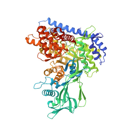
    5OAS

  • PubMed Abstract: 

    Pseudomonas aeruginosa is an opportunistic human pathogen recognized as a critical threat by the World Health Organization because of the dwindling number of effective therapies available to treat infections. Over the past decade, it has become apparent that the glyoxylate shunt plays a vital role in sustaining P. aeruginosa during infection scenarios. The glyoxylate shunt comprises two enzymes: isocitrate lyase and malate synthase isoform G. Inactivation of these enzymes has been reported to abolish the ability of P. aeruginosa to establish infection in a mammalian model system, yet we still lack the structural information to support drug design efforts. In this work, we describe the first X-ray crystal structure of P. aeruginosa malate synthase G in the apo form at 1.62 Å resolution. The enzyme is a monomer composed of four domains and is highly conserved with homologues found in other clinically relevant microorganisms. It is also dependent on Mg 2+ for catalysis. Metal ion binding led to a change in the intrinsic fluorescence of the protein, allowing us to quantitate its affinity for Mg 2+ . We also identified putative drug binding sites in malate synthase G using computational analysis and, because of the high resolution of the experimental data, were further able to characterize its hydration properties. Our data reveal two promising binding pockets in malate synthase G that may be exploited for drug design.


  • Organizational Affiliation

    Department of Biochemistry, University of Cambridge , Cambridge CB2 1QW, U.K.


Macromolecules
Find similar proteins by:  (by identity cutoff)  |  3D Structure
Entity ID: 1
MoleculeChains Sequence LengthOrganismDetailsImage
Malate synthase G728Pseudomonas aeruginosa PAO1Mutation(s): 0 
Gene Names: glcBPA0482
EC: 2.3.3.9
UniProt
Find proteins for Q9I636 (Pseudomonas aeruginosa (strain ATCC 15692 / DSM 22644 / CIP 104116 / JCM 14847 / LMG 12228 / 1C / PRS 101 / PAO1))
Explore Q9I636 
Go to UniProtKB:  Q9I636
Entity Groups  
Sequence Clusters30% Identity50% Identity70% Identity90% Identity95% Identity100% Identity
UniProt GroupQ9I636
Sequence Annotations
Expand
  • Reference Sequence
Small Molecules
Ligands 3 Unique
IDChains Name / Formula / InChI Key2D Diagram3D Interactions
GOL
Query on GOL

Download Ideal Coordinates CCD File 
B [auth A],
C [auth A],
D [auth A],
E [auth A]
GLYCEROL
C3 H8 O3
PEDCQBHIVMGVHV-UHFFFAOYSA-N
EDO
Query on EDO

Download Ideal Coordinates CCD File 
F [auth A]
G [auth A]
H [auth A]
I [auth A]
J [auth A]
F [auth A],
G [auth A],
H [auth A],
I [auth A],
J [auth A],
K [auth A],
L [auth A],
M [auth A],
N [auth A],
O [auth A],
P [auth A],
Q [auth A],
R [auth A],
S [auth A],
T [auth A]
1,2-ETHANEDIOL
C2 H6 O2
LYCAIKOWRPUZTN-UHFFFAOYSA-N
MG
Query on MG

Download Ideal Coordinates CCD File 
U [auth A],
V [auth A]
MAGNESIUM ION
Mg
JLVVSXFLKOJNIY-UHFFFAOYSA-N
Experimental Data & Validation

Experimental Data

  • Method: X-RAY DIFFRACTION
  • Resolution: 1.62 Å
  • R-Value Free: 0.193 
  • R-Value Work: 0.167 
  • R-Value Observed: 0.168 
  • Space Group: P 21 21 21
Unit Cell:
Length ( Å )Angle ( ˚ )
a = 71.171α = 90
b = 81.197β = 90
c = 137.574γ = 90
Software Package:
Software NamePurpose
PHENIXrefinement
PHASERphasing

Structure Validation

View Full Validation Report


Entry History & Funding Information

Deposition Data


Funding OrganizationLocationGrant Number
European CommissionUnited Kingdom624620

Revision History  (Full details and data files)

  • Version 1.0: 2017-10-18
    Type: Initial release
  • Version 1.1: 2017-10-25
    Changes: Database references
  • Version 1.2: 2018-12-19
    Changes: Advisory, Data collection, Derived calculations
  • Version 1.3: 2019-10-16
    Changes: Data collection
  • Version 1.4: 2024-01-17
    Changes: Data collection, Database references, Refinement description