5NNP

Structure of Naa15/Naa10 bound to HypK-THB


Experimental Data Snapshot

  • Method: X-RAY DIFFRACTION
  • Resolution: 2.60 Å
  • R-Value Free: 0.220 
  • R-Value Work: 0.161 
  • R-Value Observed: 0.164 

wwPDB Validation   3D Report Full Report


Ligand Structure Quality Assessment 


This is version 1.0 of the entry. See complete history


Literature

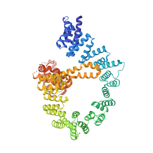
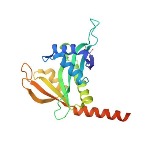
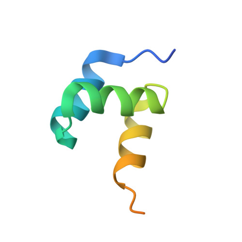
Structural basis of HypK regulating N-terminal acetylation by the NatA complex.

Weyer, F.A.Gumiero, A.Lapouge, K.Bange, G.Kopp, J.Sinning, I.

(2017) Nat Commun 8: 15726-15726

  • DOI: https://doi.org/10.1038/ncomms15726
  • Primary Citation of Related Structures:  
    5NNP, 5NNR

  • PubMed Abstract: 

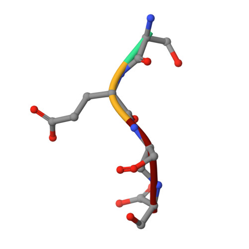
    In eukaryotes, N-terminal acetylation is one of the most common protein modifications involved in a wide range of biological processes. Most N-acetyltransferase complexes (NATs) act co-translationally, with the heterodimeric NatA complex modifying the majority of substrate proteins. Here we show that the Huntingtin yeast two-hybrid protein K (HypK) binds tightly to the NatA complex comprising the auxiliary subunit Naa15 and the catalytic subunit Naa10. The crystal structures of NatA bound to HypK or to a N-terminal deletion variant of HypK were determined without or with a bi-substrate analogue, respectively. The HypK C-terminal region is responsible for high-affinity interaction with the C-terminal part of Naa15. In combination with acetylation assays, the HypK N-terminal region is identified as a negative regulator of the NatA acetylation activity. Our study provides mechanistic insights into the regulation of this pivotal protein modification.


  • Organizational Affiliation

    Heidelberg University Biochemistry Center (BZH), INF328, D-69120 Heidelberg, Germany.


Macromolecules
Find similar proteins by:  (by identity cutoff)  |  3D Structure
Entity ID: 1
MoleculeChains Sequence LengthOrganismDetailsImage
N-terminal acetyltransferase-like proteinA,
D [auth E]
745Thermochaetoides thermophila DSM 1495Mutation(s): 0 
Gene Names: CTHT_0031530
UniProt
Find proteins for G0S4M4 (Chaetomium thermophilum (strain DSM 1495 / CBS 144.50 / IMI 039719))
Explore G0S4M4 
Go to UniProtKB:  G0S4M4
Entity Groups  
Sequence Clusters30% Identity50% Identity70% Identity90% Identity95% Identity100% Identity
UniProt GroupG0S4M4
Sequence Annotations
Expand
  • Reference Sequence
Find similar proteins by:  (by identity cutoff)  |  3D Structure
Entity ID: 2
MoleculeChains Sequence LengthOrganismDetailsImage
Putative uncharacterized proteinB,
E [auth F]
195Thermochaetoides thermophila DSM 1495Mutation(s): 0 
Gene Names: CTHT_0063490
UniProt
Find proteins for G0SEE8 (Chaetomium thermophilum (strain DSM 1495 / CBS 144.50 / IMI 039719))
Explore G0SEE8 
Go to UniProtKB:  G0SEE8
Entity Groups  
Sequence Clusters30% Identity50% Identity70% Identity90% Identity95% Identity100% Identity
UniProt GroupG0SEE8
Sequence Annotations
Expand
  • Reference Sequence
Find similar proteins by:  (by identity cutoff)  |  3D Structure
Entity ID: 3
MoleculeChains Sequence LengthOrganismDetailsImage
Putative uncharacterized proteinC,
F [auth G]
62Thermochaetoides thermophila DSM 1495Mutation(s): 0 
Gene Names: CTHT_0058830
UniProt
Find proteins for G0SCY6 (Chaetomium thermophilum (strain DSM 1495 / CBS 144.50 / IMI 039719))
Explore G0SCY6 
Go to UniProtKB:  G0SCY6
Entity Groups  
Sequence Clusters30% Identity50% Identity70% Identity90% Identity95% Identity100% Identity
UniProt GroupG0SCY6
Sequence Annotations
Expand
  • Reference Sequence

Find similar proteins by:  Sequence   |   3D Structure  

Entity ID: 4
MoleculeChains Sequence LengthOrganismDetailsImage
Ser-Glu-Ser-SerG [auth I],
H [auth L]
4synthetic constructMutation(s): 0 
Sequence Annotations
Expand
  • Reference Sequence
Small Molecules
Ligands 3 Unique
IDChains Name / Formula / InChI Key2D Diagram3D Interactions
CMC
Query on CMC

Download Ideal Coordinates CCD File 
NA [auth I],
OA [auth L]
CARBOXYMETHYL COENZYME *A
C23 H38 N7 O18 P3 S
OBUOSIHPWVNVJN-GRFIIANRSA-N
PO4
Query on PO4

Download Ideal Coordinates CCD File 
EA [auth E],
FA [auth E],
MA [auth F],
U [auth A]
PHOSPHATE ION
O4 P
NBIIXXVUZAFLBC-UHFFFAOYSA-K
GOL
Query on GOL

Download Ideal Coordinates CCD File 
AA [auth E]
BA [auth E]
CA [auth E]
DA [auth E]
GA [auth F]
AA [auth E],
BA [auth E],
CA [auth E],
DA [auth E],
GA [auth F],
HA [auth F],
I [auth A],
IA [auth F],
J [auth A],
JA [auth F],
K [auth A],
KA [auth F],
L [auth A],
LA [auth F],
M [auth A],
N [auth A],
O [auth A],
P [auth A],
Q [auth A],
R [auth A],
S [auth A],
T [auth A],
V [auth B],
W [auth C],
X [auth C],
Y [auth E],
Z [auth E]
GLYCEROL
C3 H8 O3
PEDCQBHIVMGVHV-UHFFFAOYSA-N
Modified Residues  1 Unique
IDChains TypeFormula2D DiagramParent
FME
Query on FME
B,
E [auth F]
L-PEPTIDE LINKINGC6 H11 N O3 SMET
Experimental Data & Validation

Experimental Data

  • Method: X-RAY DIFFRACTION
  • Resolution: 2.60 Å
  • R-Value Free: 0.220 
  • R-Value Work: 0.161 
  • R-Value Observed: 0.164 
  • Space Group: P 1 21 1
Unit Cell:
Length ( Å )Angle ( ˚ )
a = 93.319α = 90
b = 106.041β = 94.98
c = 130.937γ = 90
Software Package:
Software NamePurpose
PHENIXrefinement
XDSdata reduction
Aimlessdata scaling
PHASERphasing

Structure Validation

View Full Validation Report


Ligand Structure Quality Assessment 


Entry History & Funding Information

Deposition Data


Funding OrganizationLocationGrant Number
DFGGermanyFOR967
DFGGermanyGRK1188
DFGGermanyLeibniz Program

Revision History  (Full details and data files)

  • Version 1.0: 2017-06-14
    Type: Initial release