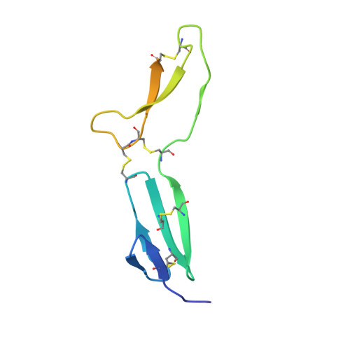
Structural analyses of von Willebrand factor C domains of collagen 2A and CCN3 reveal an alternative mode of binding to bone morphogenetic protein-2.
Xu, E.R., Blythe, E.E., Fischer, G., Hyvonen, M.(2017) J Biol Chem 292: 12516-12527
- PubMed: 28584056
- DOI: https://doi.org/10.1074/jbc.M117.788992
- Primary Citation of Related Structures:
5NB8, 5NIR - PubMed Abstract:
Bone morphogenetic proteins (BMPs) are secreted growth factors that promote differentiation processes in embryogenesis and tissue development. Regulation of BMP signaling involves binding to a variety of extracellular proteins, among which are many von Willebrand factor C (vWC) domain-containing proteins. Although the crystal structure of the complex of crossveinless-2 (CV-2) vWC1 and BMP-2 previously revealed one mode of the vWC/BMP-binding mechanism, other vWC domains may bind to BMP differently. Here, using X-ray crystallography, we present for the first time structures of the vWC domains of two proteins thought to interact with BMP-2: collagen IIA and matricellular protein CCN3. We found that these two vWC domains share a similar N-terminal fold that differs greatly from that in CV-2 vWC, which comprises its BMP-2-binding site. We analyzed the ability of these vWC domains to directly bind to BMP-2 and detected an interaction only between the collagen IIa vWC and BMP-2. Guided by the collagen IIa vWC domain crystal structure and conservation of surface residues among orthologous domains, we mapped the BMP-binding epitope on the subdomain 1 of the vWC domain. This binding site is different from that previously observed in the complex between CV-2 vWC and BMP-2, revealing an alternative mode of interaction between vWC domains and BMPs.
Organizational Affiliation:
Department of Biochemistry, University of Cambridge, Cambridge CB2 1GA, United Kingdom.