5N7W

Computationally designed functional antibody


Experimental Data Snapshot

  • Method: X-RAY DIFFRACTION
  • Resolution: 1.96 Å
  • R-Value Free: 0.250 
  • R-Value Work: 0.199 
  • R-Value Observed: 0.202 

wwPDB Validation   3D Report Full Report


This is version 1.2 of the entry. See complete history


Literature

Computational Design of Epitope-Specific Functional Antibodies.

Nimrod, G.Fischman, S.Austin, M.Herman, A.Keyes, F.Leiderman, O.Hargreaves, D.Strajbl, M.Breed, J.Klompus, S.Minton, K.Spooner, J.Buchanan, A.Vaughan, T.J.Ofran, Y.

(2018) Cell Rep 25: 2121-2131.e5

  • DOI: https://doi.org/10.1016/j.celrep.2018.10.081
  • Primary Citation of Related Structures:  
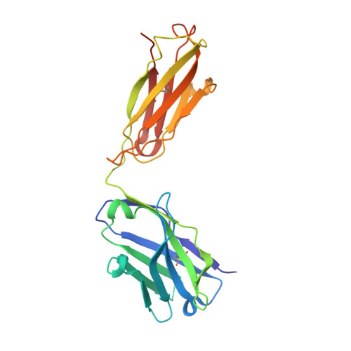
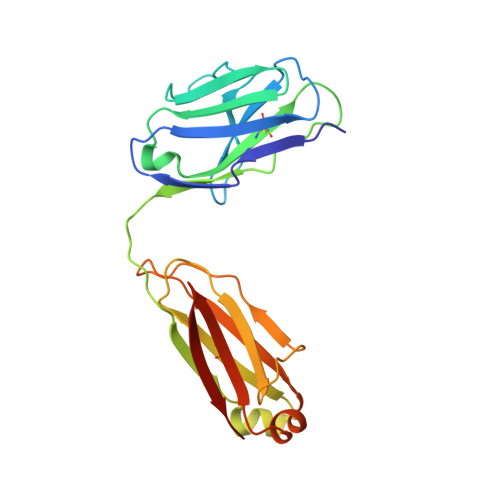
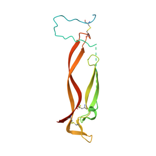
    5N7W

  • PubMed Abstract: 

    The ultimate goal of protein design is to introduce new biological activity. We propose a computational approach for designing functional antibodies by focusing on functional epitopes, integrating large-scale statistical analysis with multiple structural models. Machine learning is used to analyze these models and predict specific residue-residue contacts. We use this approach to design a functional antibody to counter the proinflammatory effect of the cytokine interleukin-17A (IL-17A). X-ray crystallography confirms that the designed antibody binds the targeted epitope and the interaction is mediated by the designed contacts. Cell-based assays confirm that the antibody is functional. Importantly, this approach does not rely on a high-quality 3D model of the designed complex or even a solved structure of the target. As demonstrated here, this approach can be used to design biologically active antibodies, removing some of the main hurdles in antibody design and in drug discovery.


  • Organizational Affiliation

    Biolojic Design, Ltd., 12 Hamada Street, Rehovot 7670314, Israel.


Macromolecules
Find similar proteins by:  (by identity cutoff)  |  3D Structure
Entity ID: 1
MoleculeChains Sequence LengthOrganismDetailsImage
Antibody Fragment Heavy ChainA,
C [auth H]
224Homo sapiensMutation(s): 0 
Entity Groups  
Sequence Clusters30% Identity50% Identity70% Identity90% Identity95% Identity100% Identity
Sequence Annotations
Expand
  • Reference Sequence
Find similar proteins by:  (by identity cutoff)  |  3D Structure
Entity ID: 2
MoleculeChains Sequence LengthOrganismDetailsImage
Antibody Fragment Light ChainB,
D [auth L]
214Homo sapiensMutation(s): 0 
Entity Groups  
Sequence Clusters30% Identity50% Identity70% Identity90% Identity95% Identity100% Identity
Sequence Annotations
Expand
  • Reference Sequence
Find similar proteins by:  (by identity cutoff)  |  3D Structure
Entity ID: 3
MoleculeChains Sequence LengthOrganismDetailsImage
Interleukin-17AE [auth X],
F [auth Y]
155Homo sapiensMutation(s): 0 
Gene Names: IL17ACTLA8IL17
UniProt & NIH Common Fund Data Resources
Find proteins for Q16552 (Homo sapiens)
Explore Q16552 
Go to UniProtKB:  Q16552
PHAROS:  Q16552
GTEx:  ENSG00000112115 
Entity Groups  
Sequence Clusters30% Identity50% Identity70% Identity90% Identity95% Identity100% Identity
UniProt GroupQ16552
Sequence Annotations
Expand
  • Reference Sequence
Experimental Data & Validation

Experimental Data

  • Method: X-RAY DIFFRACTION
  • Resolution: 1.96 Å
  • R-Value Free: 0.250 
  • R-Value Work: 0.199 
  • R-Value Observed: 0.202 
  • Space Group: P 1 21 1
Unit Cell:
Length ( Å )Angle ( ˚ )
a = 42.24α = 90
b = 199.98β = 96.42
c = 76.35γ = 90
Software Package:
Software NamePurpose
REFMACrefinement
xia2data reduction
Aimlessdata scaling
PHASERphasing

Structure Validation

View Full Validation Report


Entry History 

Deposition Data

Revision History  (Full details and data files)

  • Version 1.0: 2018-11-14
    Type: Initial release
  • Version 1.1: 2018-11-21
    Changes: Data collection, Database references
  • Version 1.2: 2018-12-05
    Changes: Data collection, Database references