5N4J

human Fab fragment 10C3 against NHBA from Neisseria meningitidis


Experimental Data Snapshot

  • Method: X-RAY DIFFRACTION
  • Resolution: 1.50 Å
  • R-Value Free: 0.211 
  • R-Value Work: 0.179 
  • R-Value Observed: 0.180 

wwPDB Validation   3D Report Full Report


This is version 1.4 of the entry. See complete history


Literature

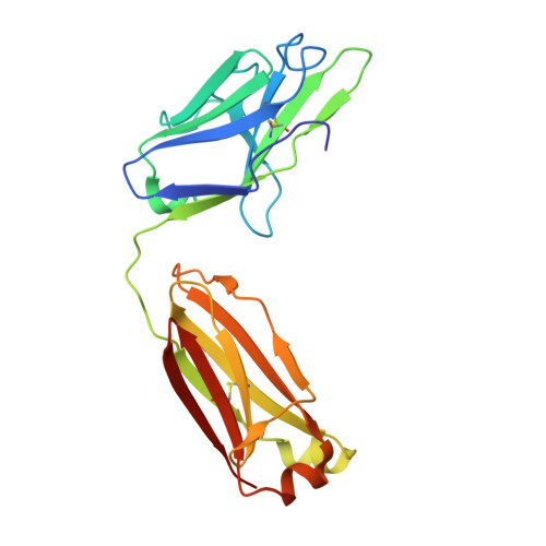
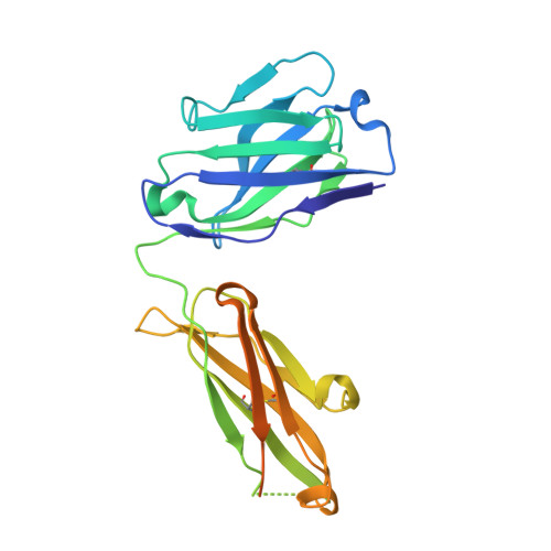
Crystal structures of human Fabs targeting the Bexsero meningococcal vaccine antigen NHBA.

Maritan, M.Cozzi, R.Lo Surdo, P.Veggi, D.Bottomley, M.J.Malito, E.

(2017) Acta Crystallogr F Struct Biol Commun 73: 305-314

  • DOI: https://doi.org/10.1107/S2053230X17006021
  • Primary Citation of Related Structures:  
    5N4G, 5N4J

  • PubMed Abstract: 

    Neisserial heparin-binding antigen (NHBA) is a surface-exposed lipoprotein from Neisseria meningitidis and is a component of the meningococcus B vaccine Bexsero. As part of a study to characterize the three-dimensional structure of NHBA and the molecular basis of the human immune response to Bexsero, the crystal structures of two fragment antigen-binding domains (Fabs) isolated from human monoclonal antibodies targeting NHBA were determined. Through a high-resolution analysis of the organization and the amino-acid composition of the CDRs, these structures provide broad insights into the NHBA epitopes recognized by the human immune system. As expected, these Fabs also show remarkable structural conservation, as shown by a structural comparison of 15 structures of apo Fab 10C3 which were obtained from crystals grown in different crystallization conditions and were solved while searching for a complex with a bound NHBA fragment or epitope peptide. This study also provides indirect evidence for the intrinsically disordered nature of two N-terminal regions of NHBA.


  • Organizational Affiliation

    GSK Vaccines, Via Fiorentina 1, 53100 Siena, Italy.


Macromolecules
Find similar proteins by:  (by identity cutoff)  |  3D Structure
Entity ID: 1
MoleculeChains Sequence LengthOrganismDetailsImage
LIGHT CHAINA [auth L]216Homo sapiensMutation(s): 0 
Entity Groups  
Sequence Clusters30% Identity50% Identity70% Identity90% Identity95% Identity100% Identity
Sequence Annotations
Expand
  • Reference Sequence
Find similar proteins by:  (by identity cutoff)  |  3D Structure
Entity ID: 2
MoleculeChains Sequence LengthOrganismDetailsImage
HEAVY CHAINB [auth H]257Homo sapiensMutation(s): 0 
Entity Groups  
Sequence Clusters30% Identity50% Identity70% Identity90% Identity95% Identity100% Identity
Sequence Annotations
Expand
  • Reference Sequence
Experimental Data & Validation

Experimental Data

  • Method: X-RAY DIFFRACTION
  • Resolution: 1.50 Å
  • R-Value Free: 0.211 
  • R-Value Work: 0.179 
  • R-Value Observed: 0.180 
  • Space Group: P 21 21 21
Unit Cell:
Length ( Å )Angle ( ˚ )
a = 69.91α = 90
b = 79.83β = 90
c = 82.58γ = 90
Software Package:
Software NamePurpose
PHENIXrefinement
XDSdata reduction
XSCALEdata scaling
PHASERphasing

Structure Validation

View Full Validation Report


Entry History 

Deposition Data

Revision History  (Full details and data files)

  • Version 1.0: 2017-05-31
    Type: Initial release
  • Version 1.1: 2017-06-14
    Changes: Database references
  • Version 1.2: 2018-01-31
    Changes: Database references
  • Version 1.3: 2019-04-03
    Changes: Data collection, Source and taxonomy
  • Version 1.4: 2024-01-17
    Changes: Data collection, Database references, Refinement description