5MT0

COMPLEMENT FACTOR D IN COMPLEX WITH A REVERSIBLE INDOLE CARBOXYLIC ACID BASED INHIBITOR


Experimental Data Snapshot

  • Method: X-RAY DIFFRACTION
  • Resolution: 1.29 Å
  • R-Value Free: 0.196 
  • R-Value Work: 0.154 

wwPDB Validation   3D Report Full Report


Ligand Structure Quality Assessment 


This is version 1.2 of the entry. See complete history


Literature

Structure-Based Library Design and Fragment Screening for the Identification of Reversible Complement Factor D Protease Inhibitors.

Vulpetti, A.Randl, S.Rudisser, S.Ostermann, N.Erbel, P.Mac Sweeney, A.Zoller, T.Salem, B.Gerhartz, B.Cumin, F.Hommel, U.Dalvit, C.Lorthiois, E.Maibaum, J.

(2017) J Med Chem 60: 1946-1958

  • DOI: https://doi.org/10.1021/acs.jmedchem.6b01684
  • Primary Citation of Related Structures:  
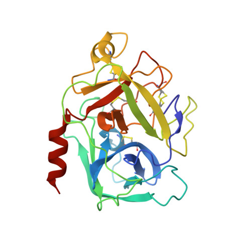
    5MT0, 5MT4

  • PubMed Abstract: 

    Chronic dysregulation of alternative complement pathway activation has been associated with diverse clinical disorders including age-related macular degeneration and paroxysmal nocturnal hemoglobinurea. Factor D is a trypsin-like serine protease with a narrow specificity for arginine in the P1 position, which catalyzes the first enzymatic reaction of the amplification loop of the alternative pathway. In this article, we describe two hit finding approaches leading to the discovery of new chemical matter for this pivotal protease of the complement system: in silico active site mapping for hot spot identification to guide rational structure-based design and NMR screening of focused and diverse fragment libraries. The wealth of information gathered by these complementary approaches enabled the identification of ligands binding to different subpockets of the latent Factor D conformation and was instrumental for understanding the binding requirements for the generation of the first known potent noncovalent reversible Factor D inhibitors.


  • Organizational Affiliation

    Novartis Institutes for BioMedical Research, Novartis Pharma AG , Novartis Campus, CH-4056 Basel, Switzerland.


Macromolecules
Find similar proteins by:  (by identity cutoff)  |  3D Structure
Entity ID: 1
MoleculeChains Sequence LengthOrganismDetailsImage
Complement factor D232Homo sapiensMutation(s): 0 
Gene Names: CFDDFPFD
EC: 3.4.21.46
UniProt & NIH Common Fund Data Resources
Find proteins for P00746 (Homo sapiens)
Explore P00746 
Go to UniProtKB:  P00746
PHAROS:  P00746
GTEx:  ENSG00000197766 
Entity Groups  
Sequence Clusters30% Identity50% Identity70% Identity90% Identity95% Identity100% Identity
UniProt GroupP00746
Sequence Annotations
Expand
  • Reference Sequence
Small Molecules
Ligands 2 Unique
IDChains Name / Formula / InChI Key2D Diagram3D Interactions
QJS
Query on QJS

Download Ideal Coordinates CCD File 
B [auth A]5-fluoranyl-3-[[(1~{S},2~{S})-2-phenylcyclopropyl]carbonylamino]-1~{H}-indole-2-carboxylic acid
C19 H15 F N2 O3
LBCNHUJFDVBZDB-OLZOCXBDSA-N
SO4
Query on SO4

Download Ideal Coordinates CCD File 
C [auth A]SULFATE ION
O4 S
QAOWNCQODCNURD-UHFFFAOYSA-L
Binding Affinity Annotations 
IDSourceBinding Affinity
QJS BindingDB:  5MT0 Ki: 5.00e+5 (nM) from 1 assay(s)
Kd: 5.00e+5 (nM) from 1 assay(s)
Binding MOAD:  5MT0 Kd: 5.00e+5 (nM) from 1 assay(s)
Experimental Data & Validation

Experimental Data

  • Method: X-RAY DIFFRACTION
  • Resolution: 1.29 Å
  • R-Value Free: 0.196 
  • R-Value Work: 0.154 
  • Space Group: P 1 21 1
Unit Cell:
Length ( Å )Angle ( ˚ )
a = 55.477α = 90
b = 49.527β = 106.36
c = 39.263γ = 90
Software Package:
Software NamePurpose
XDSdata reduction
XSCALEdata scaling
REFMACphasing
REFMACrefinement

Structure Validation

View Full Validation Report


Ligand Structure Quality Assessment 


Entry History 

Deposition Data

Revision History  (Full details and data files)

  • Version 1.0: 2017-02-15
    Type: Initial release
  • Version 1.1: 2017-03-22
    Changes: Database references
  • Version 1.2: 2019-02-20
    Changes: Advisory, Data collection, Derived calculations