Structural bases of the altered catalytic properties of a pathogenic variant of apoptosis inducing factor.
Sorrentino, L., Cossu, F., Milani, M., Aliverti, A., Mastrangelo, E.(2017) Biochem Biophys Res Commun 490: 1011-1017
- PubMed: 28666871
- DOI: https://doi.org/10.1016/j.bbrc.2017.06.156
- Primary Citation of Related Structures:
5MIU, 5MIV - PubMed Abstract:
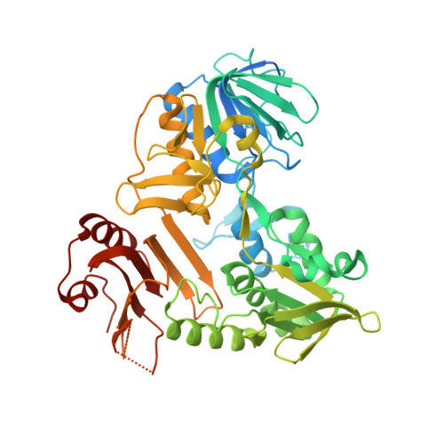
The apoptosis-inducing factor (AIF) is a FAD-containing protein playing critical roles in caspase-independent apoptosis and mitochondrial respiratory chain biogenesis and maintenance. While its lethal role is well known, the details of its mitochondrial function remain elusive. So far, nineteen allelic variants of AIF have been associated to human diseases, mainly affecting the nervous system. A strict correlation is emerging between the degree of impairment of its ability to stabilize the charge-transfer (CT) complex between FAD and NAD + and the severity of the resulting pathology. Recently, we demonstrated that the G307E replacement in murine AIF (equivalent to the pathogenic G308E in the human protein) dramatically decreases the rate of CT complex formation through the destabilization of the flavoprotein interaction with NAD(H). To provide further insights into the structural bases of its altered functional properties, here we report the first crystal structure of an AIF pathogenic mutant variant in complex with NAD + (murine AIF-G307E CT ) in comparison with its oxidized form. With respect to wild type AIF, the mutation leads to an altered positioning of NAD + adenylate moiety, which slows down CT complex formation. Moreover, the altered balance between the binding of the adenine/nicotinamide portions of the coenzyme determines a large drop in AIF-G307E ability to discriminate between NADH and NADPH.
Organizational Affiliation:
Biophysics Institute, National Research Council c/o Department of Biosciences, Università degli Studi di Milano, Via Celoria 26, 20133 Milano, Italy; Department of Biosciences, Università degli Studi di Milano, Via Celoria 26, 20133 Milano, Italy.