Structural studies of substrate and product complexes of 5-aminolaevulinic acid dehydratase from humans, Escherichia coli and the hyperthermophile Pyrobaculum calidifontis.
Mills-Davies, N., Butler, D., Norton, E., Thompson, D., Sarwar, M., Guo, J., Gill, R., Azim, N., Coker, A., Wood, S.P., Erskine, P.T., Coates, L., Cooper, J.B., Rashid, N., Akhtar, M., Shoolingin-Jordan, P.M.(2017) Acta Crystallogr D Struct Biol 73: 9-21
- PubMed: 28045381
- DOI: https://doi.org/10.1107/S2059798316019525
- Primary Citation of Related Structures:
5HMS, 5HNR, 5LZL, 5MHB - PubMed Abstract:
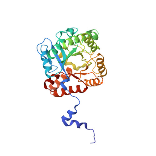
A number of X-ray analyses of an enzyme involved in a key early stage of tetrapyrrole biosynthesis are reported. Two structures of human 5-aminolaevulinate dehydratase (ALAD), native and recombinant, have been determined at 2.8 Å resolution, showing that the enzyme adopts an octameric quaternary structure in accord with previously published analyses of the enzyme from a range of other species. However, this is in contrast to the finding that a disease-related F12L mutant of the human enzyme uniquely forms hexamers [Breinig et al. (2003), Nature Struct. Biol. 10, 757-763]. Monomers of all ALADs adopt the TIM-barrel fold; the subunit conformation that assembles into the octamer includes the N-terminal tail of one monomer curled around the (α/β) 8 barrel of a neighbouring monomer. Both crystal forms of the human enzyme possess two monomers per asymmetric unit, termed A and B. In the native enzyme there are a number of distinct structural differences between the A and B monomers, with the latter exhibiting greater disorder in a number of loop regions and in the active site. In contrast, the second monomer of the recombinant enzyme appears to be better defined and the active site of both monomers clearly possesses a zinc ion which is bound by three conserved cysteine residues. In native human ALAD, the A monomer also has a ligand resembling the substrate ALA which is covalently bound by a Schiff base to one of the active-site lysines (Lys252) and is held in place by an ordered active-site loop. In contrast, these features of the active-site structure are disordered or absent in the B subunit of the native human enzyme. The octameric structure of the zinc-dependent ALAD from the hyperthermophile Pyrobaculum calidifontis is also reported at a somewhat lower resolution of 3.5 Å. Finally, the details are presented of a high-resolution structure of the Escherichia coli ALAD enzyme co-crystallized with a noncovalently bound moiety of the product, porphobilinogen (PBG). This structure reveals that the pyrrole side-chain amino group is datively bound to the active-site zinc ion and that the PBG carboxylates interact with the enzyme via hydrogen bonds and salt bridges with invariant residues. A number of hydrogen-bond interactions that were previously observed in the structure of yeast ALAD with a cyclic intermediate resembling the product PBG appear to be weaker in the new structure, suggesting that these interactions are only optimal in the transition state.
Organizational Affiliation:
School of Biological Sciences, University of Southampton, Southampton SO16 1BJ, England.