


Crystal structure of 3-Ketoacyl-CoA Thiolase (MmgA) from Bacillus subtilis.
Baker, G.E., Race, P.R.To be published.
Experimental Data Snapshot
wwPDB Validation 3D Report Full Report

Entity ID: 1 | |||||
|---|---|---|---|---|---|
| Molecule | Chains | Sequence Length | Organism | Details | Image |
| Acetyl-CoA acetyltransferase | 393 | Bacillus subtilis subsp. subtilis str. 168 | Mutation(s): 0 Gene Names: mmgA, yqiL, BSU24170 EC: 2.3.1.9 | 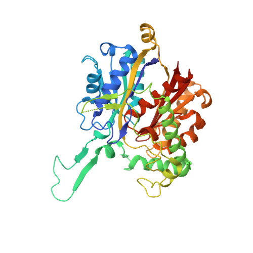 | |
UniProt | |||||
Find proteins for P45855 (Bacillus subtilis (strain 168)) Explore P45855 Go to UniProtKB: P45855 | |||||
Entity Groups | |||||
| Sequence Clusters | 30% Identity50% Identity70% Identity90% Identity95% Identity100% Identity | ||||
| UniProt Group | P45855 | ||||
Sequence AnnotationsExpand | |||||
| |||||
| Ligands 1 Unique | |||||
|---|---|---|---|---|---|
| ID | Chains | Name / Formula / InChI Key | 2D Diagram | 3D Interactions | |
| GOL Query on GOL | I [auth E] J [auth A] K [auth B] L [auth B] M [auth C] | GLYCEROL C3 H8 O3 PEDCQBHIVMGVHV-UHFFFAOYSA-N |  | ||
| Length ( Å ) | Angle ( ˚ ) |
|---|---|
| a = 55.18 | α = 90 |
| b = 141.12 | β = 89.98 |
| c = 211.462 | γ = 90 |
| Software Name | Purpose |
|---|---|
| REFMAC | refinement |
| XDS | data reduction |
| Aimless | data scaling |
| MOLREP | phasing |
RCSB PDB (citation) is hosted by
RCSB PDB is a member of the

