5LDY

Structure of Yersinia pseudotuberculosis InvD


Experimental Data Snapshot

  • Method: X-RAY DIFFRACTION
  • Resolution: 2.60 Å
  • R-Value Free: 0.253 
  • R-Value Work: 0.230 
  • R-Value Observed: 0.233 

wwPDB Validation   3D Report Full Report


This is version 1.1 of the entry. See complete history


Literature

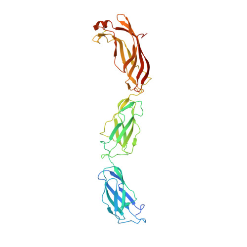
The invasin D protein from Yersinia pseudotuberculosis selectively binds the Fab region of host antibodies and affects colonization of the intestine

Sadana, P.Geyer, R.Pezoldt, J.Helmsing, S.Huehn, J.Hust, M.Dersch, P.Scrima, A.

(2018) J Biol Chem 


Macromolecules
Find similar proteins by:  (by identity cutoff)  |  3D Structure
Entity ID: 1
MoleculeChains Sequence LengthOrganismDetailsImage
Ig domain protein group 1 domain protein
A, B
357Yersinia pseudotuberculosis YPIIIMutation(s): 0 
Gene Names: YPK_1315
UniProt
Find proteins for A0A0H3B1G5 (Yersinia pseudotuberculosis serotype O:3 (strain YPIII))
Explore A0A0H3B1G5 
Go to UniProtKB:  A0A0H3B1G5
Entity Groups  
Sequence Clusters30% Identity50% Identity70% Identity90% Identity95% Identity100% Identity
UniProt GroupA0A0H3B1G5
Sequence Annotations
Expand
  • Reference Sequence
Experimental Data & Validation

Experimental Data

  • Method: X-RAY DIFFRACTION
  • Resolution: 2.60 Å
  • R-Value Free: 0.253 
  • R-Value Work: 0.230 
  • R-Value Observed: 0.233 
  • Space Group: P 43
Unit Cell:
Length ( Å )Angle ( ˚ )
a = 58.9α = 90
b = 58.9β = 90
c = 274.59γ = 90
Software Package:
Software NamePurpose
PHENIXrefinement
XSCALEdata scaling
PDB_EXTRACTdata extraction
PHENIXphasing
Cootmodel building

Structure Validation

View Full Validation Report


Entry History & Funding Information

Deposition Data


Funding OrganizationLocationGrant Number
Helmholtz AssociationGermanyVH-NG-727
Helmholtz AssociationGermanyVH-GS-202

Revision History  (Full details and data files)

  • Version 1.0: 2017-06-14
    Type: Initial release
  • Version 1.1: 2020-06-17
    Changes: Database references