5L6V

Crystal structure of E. coli ADP-glucose pyrophosphorylase (AGPase) in complex with a negative allosteric regulator adenosine monophosphate (AMP) - AGPase*AMP


Experimental Data Snapshot

  • Method: X-RAY DIFFRACTION
  • Resolution: 2.67 Å
  • R-Value Free: 0.263 
  • R-Value Work: 0.239 
  • R-Value Observed: 0.240 

wwPDB Validation   3D Report Full Report


Ligand Structure Quality Assessment 


This is version 2.1 of the entry. See complete history


Literature

Structural Basis of Glycogen Biosynthesis Regulation in Bacteria.

Cifuente, J.O.Comino, N.Madariaga-Marcos, J.Lopez-Fernandez, S.Garcia-Alija, M.Agirre, J.Albesa-Jove, D.Guerin, M.E.

(2016) Structure 24: 1613-1622

  • DOI: https://doi.org/10.1016/j.str.2016.06.023
  • Primary Citation of Related Structures:  
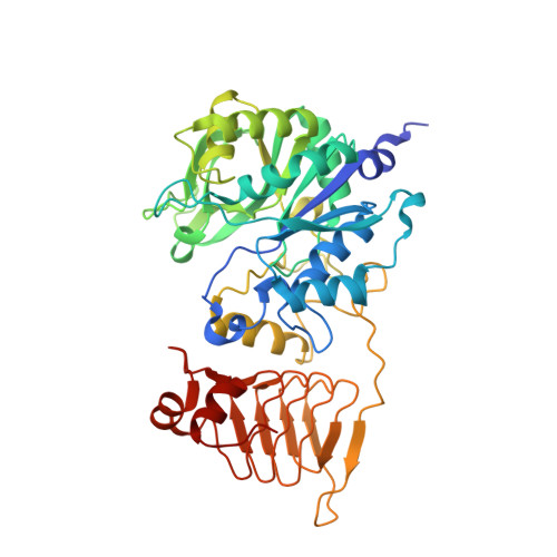
    5L6S, 5L6V

  • PubMed Abstract: 

    ADP-glucose pyrophosphorylase (AGPase) catalyzes the rate-limiting step of bacterial glycogen and plant starch biosynthesis, the most common carbon storage polysaccharides in nature. A major challenge is to understand how AGPase activity is regulated by metabolites in the energetic flux within the cell. Here we report crystal structures of the homotetrameric AGPase from Escherichia coli in complex with its physiological positive and negative allosteric regulators, fructose-1,6-bisphosphate (FBP) and AMP, and sucrose in the active site. FBP and AMP bind to partially overlapping sites located in a deep cleft between glycosyltransferase A-like and left-handed β helix domains of neighboring protomers, accounting for the fact that sensitivity to inhibition by AMP is modulated by the concentration of the activator FBP. We propose a model in which the energy reporters regulate EcAGPase catalytic activity by intra-protomer interactions and inter-protomer crosstalk, with a sensory motif and two regulatory loops playing a prominent role.


  • Organizational Affiliation

    Structural Biology Unit, CIC bioGUNE, Bizkaia Technology Park, 48160 Derio, Spain; Unidad de Biofísica, Centro Mixto Consejo Superior de Investigaciones Científicas, Universidad del País Vasco/Euskal Herriko Unibertsitatea (CSIC,UPV/EHU), Barrio Sarriena s/n, Leioa, Bizkaia, 48940, Spain; Departamento de Bioquímica, Universidad del País Vasco, 48940 Leioa, Bizkaia, Spain.


Macromolecules
Find similar proteins by:  (by identity cutoff)  |  3D Structure
Entity ID: 1
MoleculeChains Sequence LengthOrganismDetailsImage
Glucose-1-phosphate adenylyltransferase
A, B, C, D, E
A, B, C, D, E, F, G, H, I, J, K, L, M, N, O, P
431Escherichia coli K-12Mutation(s): 0 
Gene Names: glgCb3430JW3393
EC: 2.7.7.27
UniProt
Find proteins for P0A6V1 (Escherichia coli (strain K12))
Explore P0A6V1 
Go to UniProtKB:  P0A6V1
Entity Groups  
Sequence Clusters30% Identity50% Identity70% Identity90% Identity95% Identity100% Identity
UniProt GroupP0A6V1
Sequence Annotations
Expand
  • Reference Sequence
Oligosaccharides

Help

Entity ID: 2
MoleculeChains Length2D Diagram Glycosylation3D Interactions
beta-D-fructofuranose-(2-1)-alpha-D-glucopyranose
Q, R, S, T, U
Q, R, S, T, U, V, W, X
2N/A
Glycosylation Resources
GlyTouCan:  G05551OP
GlyCosmos:  G05551OP
Small Molecules
Ligands 2 Unique
IDChains Name / Formula / InChI Key2D Diagram3D Interactions
AMP
Query on AMP

Download Ideal Coordinates CCD File 
AA [auth C]
BA [auth D]
DA [auth E]
EA [auth F]
FA [auth G]
AA [auth C],
BA [auth D],
DA [auth E],
EA [auth F],
FA [auth G],
GA [auth H],
HA [auth I],
IA [auth J],
JA [auth K],
KA [auth L],
LA [auth M],
MA [auth N],
NA [auth O],
OA [auth P],
Y [auth A],
Z [auth B]
ADENOSINE MONOPHOSPHATE
C10 H14 N5 O7 P
UDMBCSSLTHHNCD-KQYNXXCUSA-N
PO4
Query on PO4

Download Ideal Coordinates CCD File 
CA [auth D]PHOSPHATE ION
O4 P
NBIIXXVUZAFLBC-UHFFFAOYSA-K
Experimental Data & Validation

Experimental Data

  • Method: X-RAY DIFFRACTION
  • Resolution: 2.67 Å
  • R-Value Free: 0.263 
  • R-Value Work: 0.239 
  • R-Value Observed: 0.240 
  • Space Group: P 1 21 1
Unit Cell:
Length ( Å )Angle ( ˚ )
a = 166.883α = 90
b = 148.193β = 115.48
c = 180.164γ = 90
Software Package:
Software NamePurpose
PHENIXrefinement
XDSdata reduction
XDSdata scaling
PHASERphasing

Structure Validation

View Full Validation Report


Ligand Structure Quality Assessment 


Entry History 

Deposition Data

Revision History  (Full details and data files)

  • Version 1.0: 2016-09-07
    Type: Initial release
  • Version 1.1: 2016-09-14
    Changes: Database references
  • Version 2.0: 2020-07-29
    Type: Remediation
    Reason: Carbohydrate remediation
    Changes: Atomic model, Data collection, Derived calculations, Non-polymer description, Structure summary
  • Version 2.1: 2024-01-10
    Changes: Data collection, Database references, Refinement description, Structure summary