


5L6S
Crystal structure of E. coli ADP-glucose pyrophosphorylase (AGPase) in complex with a positive allosteric regulator beta-fructose-1,6-diphosphate (FBP) - AGPase*FBP
- PDB DOI: https://doi.org/10.2210/pdb5L6S/pdb
- Classification: TRANSFERASE
- Organism(s): Escherichia coli K-12
- Expression System: Escherichia coli BL21(DE3)
- Mutation(s): No
- Deposited: 2016-05-31 Released: 2016-09-07
Experimental Data Snapshot
- Method: X-RAY DIFFRACTION
- Resolution: 3.04 Å
- R-Value Free: 0.272
- R-Value Work: 0.234
- R-Value Observed: 0.235

This is version 1.3 of the entry. See complete history.

Macromolecules
Find similar proteins by:
(by identity cutoff) | 3D Structure
Entity ID: 1 | |||||
|---|---|---|---|---|---|
| Molecule | Chains | Sequence Length | Organism | Details | Image |
| Glucose-1-phosphate adenylyltransferase | 431 | Escherichia coli K-12 | Mutation(s): 0 Gene Names: glgC, b3430, JW3393 EC: 2.7.7.27 | 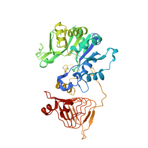 | |
UniProt | |||||
Find proteins for P0A6V1 (Escherichia coli (strain K12)) Explore P0A6V1 Go to UniProtKB: P0A6V1 | |||||
Entity Groups | |||||
| Sequence Clusters | 30% Identity50% Identity70% Identity90% Identity95% Identity100% Identity | ||||
| UniProt Group | P0A6V1 | ||||
Sequence AnnotationsExpand | |||||
| |||||
Small Molecules
| Ligands 2 Unique | |||||
|---|---|---|---|---|---|
| ID | Chains | Name / Formula / InChI Key | 2D Diagram | 3D Interactions | |
| FBP Query on FBP | EB [auth L], MA [auth G], UA [auth I], Z [auth C] | 1,6-di-O-phosphono-beta-D-fructofuranose C6 H14 O12 P2 RNBGYGVWRKECFJ-ARQDHWQXSA-N |  | ||
| SO4 Query on SO4 | AA [auth D] AB [auth K] BA [auth D] BB [auth L] CA [auth D] | SULFATE ION O4 S QAOWNCQODCNURD-UHFFFAOYSA-L |  | ||
Experimental Data & Validation
Experimental Data
- Method: X-RAY DIFFRACTION
- Resolution: 3.04 Å
- R-Value Free: 0.272
- R-Value Work: 0.234
- R-Value Observed: 0.235
- Space Group: P 1 21 1
Unit Cell:
| Length ( Å ) | Angle ( ˚ ) |
|---|---|
| a = 161.16 | α = 90 |
| b = 148.9 | β = 113.1 |
| c = 177.49 | γ = 90 |
| Software Name | Purpose |
|---|---|
| PHENIX | refinement |
| XDS | data reduction |
| XDS | data scaling |
| PHASER | phasing |
Entry History
Deposition Data
- Released Date: 2016-09-07 Deposition Author(s): Cifuente, J.O., Albesa-Jove, D., Comino, N., Madariaga-Marcos, J., Agirre, J., Lopez-Fernandez, S., Garcia-Alija, M., Guerin, M.E.
Revision History (Full details and data files)
- Version 1.0: 2016-09-07
Type: Initial release - Version 1.1: 2016-09-14
Changes: Database references - Version 1.2: 2020-07-29
Type: Remediation
Reason: Carbohydrate remediation
Changes: Data collection, Derived calculations, Structure summary - Version 1.3: 2024-01-10
Changes: Data collection, Database references, Refinement description, Structure summary
