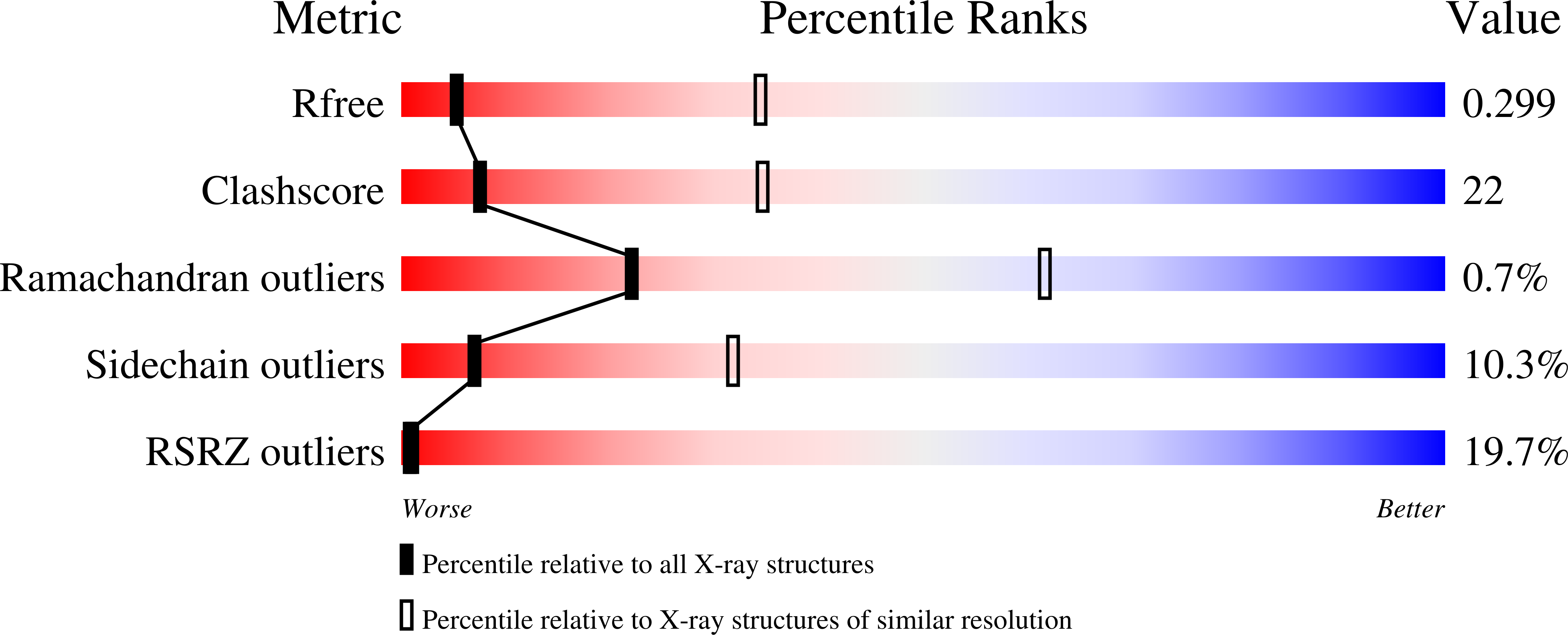
5KZF

Crystal structure of near full-length hexameric Mycobacterium tuberculosis proteasomal ATPase Mpa in apo form


Experimental Data Snapshot

  • Method: X-RAY DIFFRACTION
  • Resolution: 3.49 Å
  • R-Value Free: 0.306 
  • R-Value Work: 0.268 
  • R-Value Observed: 0.269 

wwPDB Validation   3D Report Full Report


This is version 1.4 of the entry. See complete history


Literature

Mycobacterium tuberculosis proteasomal ATPase Mpa has a beta-grasp domain that hinders docking with the proteasome core protease.

Wu, Y.Hu, K.Li, D.Bai, L.Yang, S.Jastrab, J.B.Xiao, S.Hu, Y.Zhang, S.Darwin, K.H.Wang, T.Li, H.

(2017) Mol Microbiol 105: 227-241

  • DOI: https://doi.org/10.1111/mmi.13695
  • Primary Citation of Related Structures:  
    5KWA, 5KZF

  • PubMed Abstract: 

    Mycobacterium tuberculosis (Mtb) has a proteasome system that is essential for its ability to cause lethal infections in mice. A key component of the system is the proteasomal adenosine triphosphatase (ATPase) Mpa, which captures, unfolds, and translocates protein substrates into the Mtb proteasome core particle for degradation. Here, we report the crystal structures of near full-length hexameric Mtb Mpa in apo and ADP-bound forms. Surprisingly, the structures revealed a ubiquitin-like β-grasp domain that precedes the proteasome-activating carboxyl terminus. This domain, which was only found in bacterial proteasomal ATPases, buries the carboxyl terminus of each protomer in the central channel of the hexamer and hinders the interaction of Mpa with the proteasome core protease. Thus, our work reveals the structure of a bacterial proteasomal ATPase in the hexameric form, and the structure finally explains why Mpa is unable to stimulate robust protein degradation in vitro in the absence of other, yet-to-be-identified co-factors.


  • Organizational Affiliation

    Department of Biology, Southern University of Science and Technology, 1088 Xueyuan Road, Nanshan District, Shenzhen, 518055, China.


Macromolecules
Find similar proteins by:  (by identity cutoff)  |  3D Structure
Entity ID: 1
MoleculeChains Sequence LengthOrganismDetailsImage
Proteasome-associated ATPase
A, B, C, D, E
A, B, C, D, E, F, G, H, I, J, K, L
513Mycobacterium tuberculosis H37RaMutation(s): 0 
Gene Names: mpaMRA_2130
UniProt
Find proteins for A5U4E1 (Mycobacterium tuberculosis (strain ATCC 25177 / H37Ra))
Explore A5U4E1 
Go to UniProtKB:  A5U4E1
Entity Groups  
Sequence Clusters30% Identity50% Identity70% Identity90% Identity95% Identity100% Identity
UniProt GroupA5U4E1
Sequence Annotations
Expand
  • Reference Sequence
Experimental Data & Validation

Experimental Data

  • Method: X-RAY DIFFRACTION
  • Resolution: 3.49 Å
  • R-Value Free: 0.306 
  • R-Value Work: 0.268 
  • R-Value Observed: 0.269 
  • Space Group: P 21 21 21
Unit Cell:
Length ( Å )Angle ( ˚ )
a = 115.265α = 90
b = 202.59β = 90
c = 303.007γ = 90
Software Package:
Software NamePurpose
REFMACrefinement
iMOSFLMdata reduction
SCALAdata scaling
PHASERphasing

Structure Validation

View Full Validation Report


Entry History & Funding Information

Deposition Data


Funding OrganizationLocationGrant Number
National Institutes of Health/National Institute Of Allergy and Infectious Diseases (NIH/NIAID)United StatesAI070285

Revision History  (Full details and data files)

  • Version 1.0: 2017-05-17
    Type: Initial release
  • Version 1.1: 2017-07-12
    Changes: Database references
  • Version 1.2: 2017-09-20
    Changes: Author supporting evidence
  • Version 1.3: 2019-12-11
    Changes: Advisory, Author supporting evidence, Derived calculations
  • Version 1.4: 2024-03-06
    Changes: Data collection, Database references, Refinement description