5KXV

Structure Proteinase K at 0.98 Angstroms


Experimental Data Snapshot

  • Method: X-RAY DIFFRACTION
  • Resolution: 0.98 Å
  • R-Value Free: 0.114 
  • R-Value Work: 0.100 
  • R-Value Observed: 0.101 

wwPDB Validation   3D Report Full Report


This is version 1.2 of the entry. See complete history


Literature

Atomic resolution structure of serine protease proteinase K at ambient temperature.

Masuda, T.Suzuki, M.Inoue, S.Song, C.Nakane, T.Nango, E.Tanaka, R.Tono, K.Joti, Y.Kameshima, T.Hatsui, T.Yabashi, M.Mikami, B.Nureki, O.Numata, K.Iwata, S.Sugahara, M.

(2017) Sci Rep 7: 45604-45604

  • DOI: https://doi.org/10.1038/srep45604
  • Primary Citation of Related Structures:  
    5KXU, 5KXV

  • PubMed Abstract: 

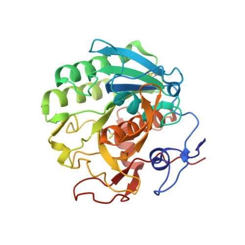
    Atomic resolution structures (beyond 1.20 Å) at ambient temperature, which is usually hampered by the radiation damage in synchrotron X-ray crystallography (SRX), will add to our understanding of the structure-function relationships of enzymes. Serial femtosecond crystallography (SFX) has attracted surging interest by providing a route to bypass such challenges. Yet the progress on atomic resolution analysis with SFX has been rather slow. In this report, we describe the 1.20 Å resolution structure of proteinase K using 13 keV photon energy. Hydrogen atoms, water molecules, and a number of alternative side-chain conformations have been resolved. The increase in the value of B-factor in SFX suggests that the residues and water molecules adjacent to active sites were flexible and exhibited dynamic motions at specific substrate-recognition sites.


  • Organizational Affiliation

    Division of Food Science and Biotechnology, Graduate School of Agriculture, Kyoto University, Gokasho, Uji, Kyoto 611-0011, Japan.


Macromolecules
Find similar proteins by:  (by identity cutoff)  |  3D Structure
Entity ID: 1
MoleculeChains Sequence LengthOrganismDetailsImage
Proteinase K279Parengyodontium albumMutation(s): 1 
EC: 3.4.21.64
UniProt
Find proteins for P06873 (Parengyodontium album)
Explore P06873 
Go to UniProtKB:  P06873
Entity Groups  
Sequence Clusters30% Identity50% Identity70% Identity90% Identity95% Identity100% Identity
UniProt GroupP06873
Sequence Annotations
Expand
  • Reference Sequence
Experimental Data & Validation

Experimental Data

  • Method: X-RAY DIFFRACTION
  • Resolution: 0.98 Å
  • R-Value Free: 0.114 
  • R-Value Work: 0.100 
  • R-Value Observed: 0.101 
  • Space Group: P 43 21 2
Unit Cell:
Length ( Å )Angle ( ˚ )
a = 67.577α = 90
b = 67.577β = 90
c = 107.14γ = 90
Software Package:
Software NamePurpose
PHENIXrefinement
HKL-2000data collection
HKL-2000data scaling
Cootmodel building

Structure Validation

View Full Validation Report


Entry History & Funding Information

Deposition Data


Funding OrganizationLocationGrant Number
Japan Science and TechnologyJapan25650026

Revision History  (Full details and data files)

  • Version 1.0: 2017-06-07
    Type: Initial release
  • Version 1.1: 2021-10-27
    Changes: Author supporting evidence, Database references, Derived calculations, Structure summary
  • Version 1.2: 2023-11-08
    Changes: Data collection, Refinement description