


Crystal structure of anthranilate synthase component I from Streptococcus pneumoniae TIGR4
Chang, C., Michalska, K., Bigelow, L., Jedrzejczak, R., ANDERSON, W.F., JOACHIMIAK, A.To be published.
Experimental Data Snapshot
wwPDB Validation 3D Report Full Report

Entity ID: 1 | |||||
|---|---|---|---|---|---|
| Molecule | Chains | Sequence Length | Organism | Details | Image |
| Anthranilate synthase component I | 450 | Streptococcus pneumoniae TIGR4 | Mutation(s): 0 Gene Names: trpE, SP_1817 EC: 4.1.3.27 | 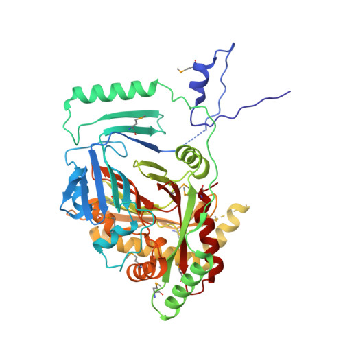 | |
UniProt | |||||
Find proteins for A0A0H2URN1 (Streptococcus pneumoniae serotype 4 (strain ATCC BAA-334 / TIGR4)) Explore A0A0H2URN1 Go to UniProtKB: A0A0H2URN1 | |||||
Entity Groups | |||||
| Sequence Clusters | 30% Identity50% Identity70% Identity90% Identity95% Identity100% Identity | ||||
| UniProt Group | A0A0H2URN1 | ||||
Sequence AnnotationsExpand | |||||
| |||||
| Ligands 1 Unique | |||||
|---|---|---|---|---|---|
| ID | Chains | Name / Formula / InChI Key | 2D Diagram | 3D Interactions | |
| GOL Query on GOL | B [auth A], C [auth A] | GLYCEROL C3 H8 O3 PEDCQBHIVMGVHV-UHFFFAOYSA-N |  | ||
| Modified Residues 1 Unique | |||||
|---|---|---|---|---|---|
| ID | Chains | Type | Formula | 2D Diagram | Parent |
| MSE Query on MSE | A | L-PEPTIDE LINKING | C5 H11 N O2 Se |  | MET |
| Length ( Å ) | Angle ( ˚ ) |
|---|---|
| a = 120.946 | α = 90 |
| b = 86.127 | β = 93.45 |
| c = 55.792 | γ = 90 |
| Software Name | Purpose |
|---|---|
| PHENIX | refinement |
| SCALEPACK | data scaling |
| PDB_EXTRACT | data extraction |
| HKL-3000 | data reduction |
| HKL-3000 | phasing |
RCSB PDB (citation) is hosted by
RCSB PDB is a member of the

