5K59

Crystal structure of LukGH from Staphylococcus aureus in complex with a neutralising antibody


Experimental Data Snapshot

  • Method: X-RAY DIFFRACTION
  • Resolution: 2.84 Å
  • R-Value Free: 0.237 
  • R-Value Work: 0.197 
  • R-Value Observed: 0.199 

wwPDB Validation   3D Report Full Report


This is version 1.3 of the entry. See complete history


Literature

Context matters: The importance of dimerization-induced conformation of the LukGH leukocidin of Staphylococcus aureus for the generation of neutralizing antibodies.

Badarau, A.Rouha, H.Malafa, S.Battles, M.B.Walker, L.Nielson, N.Dolezilkova, I.Teubenbacher, A.Banerjee, S.Maierhofer, B.Weber, S.Stulik, L.Logan, D.T.Welin, M.Mirkina, I.Pleban, C.Zauner, G.Gross, K.Jagerhofer, M.Magyarics, Z.Nagy, E.

(2016) MAbs 8: 1347-1360

  • DOI: https://doi.org/10.1080/19420862.2016.1215791
  • Primary Citation of Related Structures:  
    5K59

  • PubMed Abstract: 

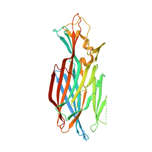
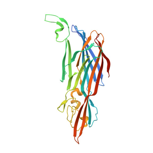
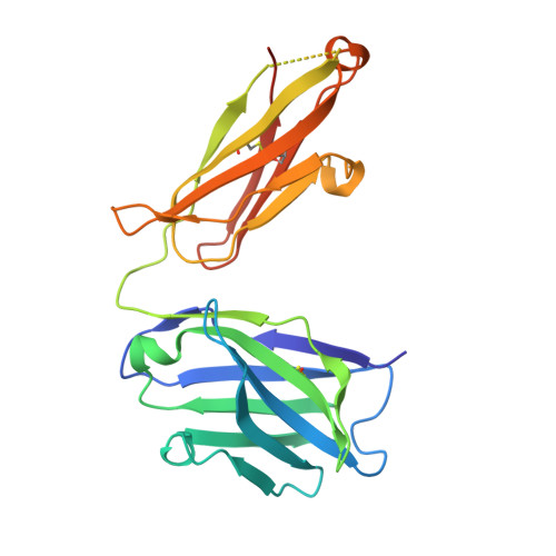
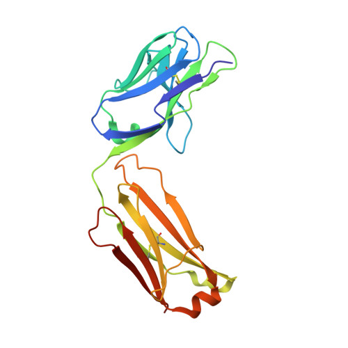
    LukGH (LukAB) is a potent leukocidin of Staphylococcus aureus that lyses human phagocytic cells and is thought to contribute to immune evasion. Unlike the other bi-component leukocidins of S. aureus, LukGH forms a heterodimer before binding to its receptor, CD11b expressed on professional phagocytic cells, and displays significant sequence variation. We employed a high diversity human IgG1 library presented on yeast cells to discover monoclonal antibodies (mAbs) neutralizing the cytolytic activity of LukGH. Recombinant LukG and LukH monomers or a LukGH dimer were used as capture antigens in the library selections. We found that mAbs identified with LukG or LukH as bait had no or very low toxin neutralization potency. In contrast, LukGH dimer-selected antibodies proved to be highly potent, and several mAbs were able to neutralize even the most divergent LukGH variants. Based on biolayer interferometry and mesoscale discovery, the high affinity antibody binding site on the LukGH complex was absent on the individual monomers, suggesting that it was generated upon formation of the LukG-LukH dimer. X-ray crystallography analysis of the complex between the LukGH dimer and the antigen-binding fragment of a very potent mAb (PDB code 5K59) indicated that the epitope is located in the predicted cell binding region (rim domain) of LukGH. The corresponding IgG inhibited the binding of LukGH dimer to target cells. Our data suggest that knowledge of the native conformation of target molecules is essential to generate high affinity and functional mAbs.


  • Organizational Affiliation

    a Arsanis Biosciences , Campus Vienna Biocenter, Vienna , Austria.


Macromolecules
Find similar proteins by:  (by identity cutoff)  |  3D Structure
Entity ID: 1
MoleculeChains Sequence LengthOrganismDetailsImage
Uncharacterized leukocidin-like protein 2A [auth D],
C
319Staphylococcus aureus subsp. aureus USA300Mutation(s): 0 
Gene Names: SAUSA300_1975
Membrane Entity: Yes 
UniProt
Find proteins for Q2FWP0 (Staphylococcus aureus (strain NCTC 8325 / PS 47))
Explore Q2FWP0 
Go to UniProtKB:  Q2FWP0
Entity Groups  
Sequence Clusters30% Identity50% Identity70% Identity90% Identity95% Identity100% Identity
UniProt GroupQ2FWP0
Sequence Annotations
Expand
  • Reference Sequence
Find similar proteins by:  (by identity cutoff)  |  3D Structure
Entity ID: 2
MoleculeChains Sequence LengthOrganismDetailsImage
Uncharacterized leukocidin-like protein 1B,
D [auth A]
311Staphylococcus aureus subsp. aureus USA300Mutation(s): 0 
Gene Names: SAUSA300_1974
Membrane Entity: Yes 
UniProt
Find proteins for Q2FFA3 (Staphylococcus aureus (strain USA300))
Explore Q2FFA3 
Go to UniProtKB:  Q2FFA3
Entity Groups  
Sequence Clusters30% Identity50% Identity70% Identity90% Identity95% Identity100% Identity
UniProt GroupQ2FFA3
Sequence Annotations
Expand
  • Reference Sequence
Find similar proteins by:  (by identity cutoff)  |  3D Structure
Entity ID: 3
MoleculeChains Sequence LengthOrganismDetailsImage
Fab heavy chainE [auth H],
G [auth E]
227Homo sapiensMutation(s): 0 
Entity Groups  
Sequence Clusters30% Identity50% Identity70% Identity90% Identity95% Identity100% Identity
Sequence Annotations
Expand
  • Reference Sequence
Find similar proteins by:  (by identity cutoff)  |  3D Structure
Entity ID: 4
MoleculeChains Sequence LengthOrganismDetailsImage
Fab light chainF [auth L],
H [auth F]
214Homo sapiensMutation(s): 0 
Entity Groups  
Sequence Clusters30% Identity50% Identity70% Identity90% Identity95% Identity100% Identity
Sequence Annotations
Expand
  • Reference Sequence
Experimental Data & Validation

Experimental Data

  • Method: X-RAY DIFFRACTION
  • Resolution: 2.84 Å
  • R-Value Free: 0.237 
  • R-Value Work: 0.197 
  • R-Value Observed: 0.199 
  • Space Group: P 1 21 1
Unit Cell:
Length ( Å )Angle ( ˚ )
a = 74.79α = 90
b = 160.932β = 101.18
c = 119.484γ = 90
Software Package:
Software NamePurpose
REFMACrefinement
XDSdata reduction
XSCALEdata scaling
PHASERphasing

Structure Validation

View Full Validation Report


Entry History 

Deposition Data

Revision History  (Full details and data files)

  • Version 1.0: 2016-08-10
    Type: Initial release
  • Version 1.1: 2016-09-14
    Changes: Structure summary
  • Version 1.2: 2016-10-12
    Changes: Database references
  • Version 1.3: 2018-01-17
    Changes: Data collection