5JYL

Human P-cadherin MEC1 with scFv TSP7 bound


Experimental Data Snapshot

  • Method: X-RAY DIFFRACTION
  • Resolution: 2.55 Å
  • R-Value Free: 0.271 
  • R-Value Work: 0.214 
  • R-Value Observed: 0.217 

wwPDB Validation   3D Report Full Report


This is version 1.3 of the entry. See complete history


Literature

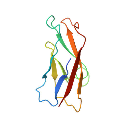
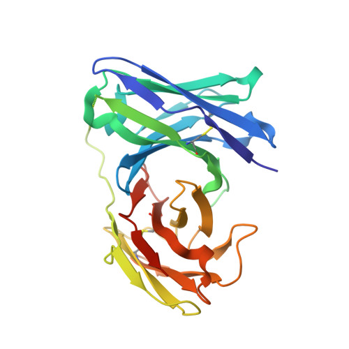
Disruption of cell adhesion by an antibody targeting the cell-adhesive intermediate (X-dimer) of human P-cadherin

Kudo, S.Caaveiro, J.M.M.Nagatoishi, S.Miyafusa, T.Matsuura, T.Sudou, Y.Tsumoto, K.

(2017) Sci Rep 7: 39518-39518

  • DOI: https://doi.org/10.1038/srep39518
  • Primary Citation of Related Structures:  
    5JYL, 5JYM

  • PubMed Abstract: 

    Human P-cadherin is a cell adhesion protein of the family of classical cadherins, the overexpression of which is correlated with poor prognosis in various types of cancer. Antibodies inhibiting cell-cell adhesion mediated by P-cadherin show clear therapeutic effect, although the mechanistic basis explaining their effectiveness is still unclear. Based on structural, physicochemical, and functional analyses, we have elucidated the molecular mechanism of disruption of cell adhesion by antibodies targeting human P-cadherin. Herein we have studied three different antibodies, TSP5, TSP7, and TSP11, each recognizing a different epitope on the surface of the cell-adhesive domain (EC1). Although all these three antibodies recognized human P-cadherin with high affinity, only TSP7 disrupted cell adhesion. Notably, we demonstrated that TSP7 abolishes cell adhesion by disabling the so-called X-dimer (a kinetic adhesive intermediate), in addition to disrupting the strand-swap dimer (the final thermodynamic state). The inhibition of the X-dimer was crucial for the overall inhibitory effect, raising the therapeutic value of a kinetic intermediary not only for preventing, but also for reversing, cell adhesion mediated by a member of the classical cadherin family. These findings should help to design more innovative and effective therapeutic solutions targeting human P-cadherin.


  • Organizational Affiliation

    Department of Chemistry &Biotechnology, School of Engineering, The University of Tokyo, Tokyo 108-8639, Japan.


Macromolecules
Find similar proteins by:  (by identity cutoff)  |  3D Structure
Entity ID: 1
MoleculeChains Sequence LengthOrganismDetailsImage
Cadherin-3
A, C
101Homo sapiensMutation(s): 0 
Gene Names: CDH3
UniProt & NIH Common Fund Data Resources
Find proteins for P22223 (Homo sapiens)
Explore P22223 
Go to UniProtKB:  P22223
PHAROS:  P22223
GTEx:  ENSG00000062038 
Entity Groups  
Sequence Clusters30% Identity50% Identity70% Identity90% Identity95% Identity100% Identity
UniProt GroupP22223
Sequence Annotations
Expand
  • Reference Sequence
Find similar proteins by:  (by identity cutoff)  |  3D Structure
Entity ID: 2
MoleculeChains Sequence LengthOrganismDetailsImage
scFv TSP7
B, D
253Mus musculusMutation(s): 0 
Entity Groups  
Sequence Clusters30% Identity50% Identity70% Identity90% Identity95% Identity100% Identity
Sequence Annotations
Expand
  • Reference Sequence
Experimental Data & Validation

Experimental Data

  • Method: X-RAY DIFFRACTION
  • Resolution: 2.55 Å
  • R-Value Free: 0.271 
  • R-Value Work: 0.214 
  • R-Value Observed: 0.217 
  • Space Group: P 1 21 1
Unit Cell:
Length ( Å )Angle ( ˚ )
a = 66.57α = 90
b = 49.41β = 101.89
c = 111.66γ = 90
Software Package:
Software NamePurpose
REFMACrefinement
MOSFLMdata reduction
SCALAdata scaling
PHASERmodel building

Structure Validation

View Full Validation Report


Entry History 

Deposition Data

Revision History  (Full details and data files)

  • Version 1.0: 2016-12-28
    Type: Initial release
  • Version 1.1: 2017-01-18
    Changes: Database references
  • Version 1.2: 2020-02-19
    Changes: Data collection
  • Version 1.3: 2023-11-08
    Changes: Data collection, Database references, Refinement description