5JQ7

Crystal structure of Ebola glycoprotein in complex with toremifene


Experimental Data Snapshot

  • Method: X-RAY DIFFRACTION
  • Resolution: 2.69 Å
  • R-Value Free: 0.245 
  • R-Value Work: 0.203 
  • R-Value Observed: 0.205 

wwPDB Validation   3D Report Full Report


Ligand Structure Quality Assessment 


This is version 2.1 of the entry. See complete history


Literature

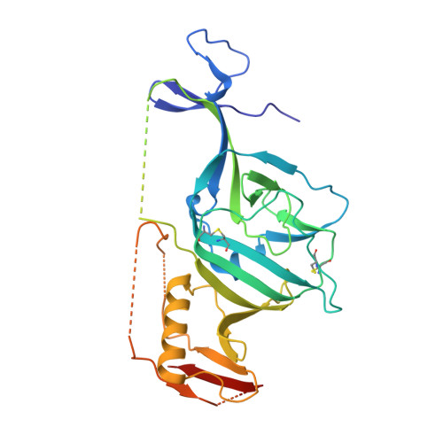
Toremifene interacts with and destabilizes the Ebola virus glycoprotein.

Zhao, Y.Ren, J.Harlos, K.Jones, D.M.Zeltina, A.Bowden, T.A.Padilla-Parra, S.Fry, E.E.Stuart, D.I.

(2016) Nature 535: 168-172

  • DOI: https://doi.org/10.1038/nature18615
  • Primary Citation of Related Structures:  
    5JQ3, 5JQ7, 5JQB

  • PubMed Abstract: 

    Ebola viruses (EBOVs) are responsible for repeated outbreaks of fatal infections, including the recent deadly epidemic in West Africa. There are currently no approved therapeutic drugs or vaccines for the disease. EBOV has a membrane envelope decorated by trimers of a glycoprotein (GP, cleaved by furin to form GP1 and GP2 subunits), which is solely responsible for host cell attachment, endosomal entry and membrane fusion. GP is thus a primary target for the development of antiviral drugs. Here we report the first, to our knowledge, unliganded structure of EBOV GP, and high-resolution complexes of GP with the anticancer drug toremifene and the painkiller ibuprofen. The high-resolution apo structure gives a more complete and accurate picture of the molecule, and allows conformational changes introduced by antibody and receptor binding to be deciphered. Unexpectedly, both toremifene and ibuprofen bind in a cavity between the attachment (GP1) and fusion (GP2) subunits at the entrance to a large tunnel that links with equivalent tunnels from the other monomers of the trimer at the three-fold axis. Protein–drug interactions with both GP1 and GP2 are predominately hydrophobic. Residues lining the binding site are highly conserved among filoviruses except Marburg virus (MARV), suggesting that MARV may not bind these drugs. Thermal shift assays show up to a 14 °C decrease in the protein melting temperature after toremifene binding, while ibuprofen has only a marginal effect and is a less potent inhibitor. These results suggest that inhibitor binding destabilizes GP and triggers premature release of GP2, thereby preventing fusion between the viral and endosome membranes. Thus, these complex structures reveal the mechanism of inhibition and may guide the development of more powerful anti-EBOV drugs.


  • Organizational Affiliation

    Division of Structural Biology, University of Oxford, The Henry Wellcome Building for Genomic Medicine, Headington, Oxford, OX3 7BN, UK.


Macromolecules
Find similar proteins by:  (by identity cutoff)  |  3D Structure
Entity ID: 1
MoleculeChains Sequence LengthOrganismDetailsImage
Envelope glycoprotein 1,Envelope glycoprotein 1,Envelope glycoprotein 1330Ebola virus - Mayinga, Zaire, 1976Mutation(s): 1 
Gene Names: GP
UniProt
Find proteins for Q05320 (Zaire ebolavirus (strain Mayinga-76))
Explore Q05320 
Go to UniProtKB:  Q05320
Entity Groups  
Sequence Clusters30% Identity50% Identity70% Identity90% Identity95% Identity100% Identity
UniProt GroupQ05320
Sequence Annotations
Expand
  • Reference Sequence
Find similar proteins by:  (by identity cutoff)  |  3D Structure
Entity ID: 2
MoleculeChains Sequence LengthOrganismDetailsImage
Envelope glycoprotein 2168Ebola virus - Mayinga, Zaire, 1976Mutation(s): 0 
Gene Names: GP
UniProt
Find proteins for Q05320 (Zaire ebolavirus (strain Mayinga-76))
Explore Q05320 
Go to UniProtKB:  Q05320
Entity Groups  
Sequence Clusters30% Identity50% Identity70% Identity90% Identity95% Identity100% Identity
UniProt GroupQ05320
Sequence Annotations
Expand
  • Reference Sequence
Oligosaccharides

Help

Entity ID: 3
MoleculeChains Length2D Diagram Glycosylation3D Interactions
alpha-D-mannopyranose-(1-6)-beta-D-mannopyranose-(1-4)-2-acetamido-2-deoxy-beta-D-glucopyranose-(1-4)-2-acetamido-2-deoxy-beta-D-glucopyranose
C
4N-Glycosylation
Glycosylation Resources
GlyTouCan:  G22573RC
GlyCosmos:  G22573RC
GlyGen:  G22573RC
Small Molecules
Ligands 4 Unique
IDChains Name / Formula / InChI Key2D Diagram3D Interactions
T0R
Query on T0R

Download Ideal Coordinates CCD File 
K [auth B] Toremifene
C26 H28 Cl N O
XFCLJVABOIYOMF-QPLCGJKRSA-N
NAG
Query on NAG

Download Ideal Coordinates CCD File 
D [auth A],
E [auth A],
F [auth A],
G [auth A]
2-acetamido-2-deoxy-beta-D-glucopyranose
C8 H15 N O6
OVRNDRQMDRJTHS-FMDGEEDCSA-N
GOL
Query on GOL

Download Ideal Coordinates CCD File 
H [auth A],
I [auth A]
GLYCEROL
C3 H8 O3
PEDCQBHIVMGVHV-UHFFFAOYSA-N
DMS
Query on DMS

Download Ideal Coordinates CCD File 
J [auth A]DIMETHYL SULFOXIDE
C2 H6 O S
IAZDPXIOMUYVGZ-UHFFFAOYSA-N
Binding Affinity Annotations 
IDSourceBinding Affinity
T0R BindingDB:  5JQ7 Kd: 1.60e+4 (nM) from 1 assay(s)
EC50: 63 (nM) from 1 assay(s)
Experimental Data & Validation

Experimental Data

  • Method: X-RAY DIFFRACTION
  • Resolution: 2.69 Å
  • R-Value Free: 0.245 
  • R-Value Work: 0.203 
  • R-Value Observed: 0.205 
  • Space Group: H 3 2
Unit Cell:
Length ( Å )Angle ( ˚ )
a = 113.45α = 90
b = 113.45β = 90
c = 306.87γ = 120
Software Package:
Software NamePurpose
REFMACrefinement
xia2data reduction
xia2data scaling
MOLREPphasing

Structure Validation

View Full Validation Report


Ligand Structure Quality Assessment 


Entry History 

Deposition Data

Revision History  (Full details and data files)

  • Version 1.0: 2016-06-29
    Type: Initial release
  • Version 1.1: 2016-07-13
    Changes: Database references
  • Version 2.0: 2020-07-29
    Type: Remediation
    Reason: Carbohydrate remediation
    Changes: Atomic model, Data collection, Derived calculations, Structure summary
  • Version 2.1: 2024-01-10
    Changes: Data collection, Database references, Derived calculations, Refinement description, Structure summary