5IW4

Crystal structure of E. coli NudC in complex with NAD


Experimental Data Snapshot

  • Method: X-RAY DIFFRACTION
  • Resolution: 2.60 Å
  • R-Value Free: 0.248 
  • R-Value Work: 0.200 
  • R-Value Observed: 0.202 

wwPDB Validation   3D Report Full Report


Ligand Structure Quality Assessment 


This is version 1.3 of the entry. See complete history


Literature

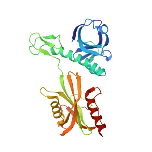
Structure and function of the bacterial decapping enzyme NudC.

Hofer, K.Li, S.Abele, F.Frindert, J.Schlotthauer, J.Grawenhoff, J.Du, J.Patel, D.J.Jaschke, A.

(2016) Nat Chem Biol 12: 730-734

  • DOI: https://doi.org/10.1038/nchembio.2132
  • Primary Citation of Related Structures:  
    5IW4, 5IW5

  • PubMed Abstract: 

    RNA capping and decapping are thought to be distinctive features of eukaryotes. The redox cofactor NAD was recently discovered to be attached to small regulatory RNAs in bacteria in a cap-like manner, and Nudix hydrolase NudC was found to act as a NAD-decapping enzyme in vitro and in vivo. Here, crystal structures of Escherichia coli NudC in complex with substrate NAD and with cleavage product NMN reveal the catalytic residues lining the binding pocket and principles underlying molecular recognition of substrate and product. Biochemical mutation analysis identifies the conserved Nudix motif as the catalytic center of the enzyme, which needs to be homodimeric, as the catalytic pocket is composed of amino acids from both monomers. NudC is single-strand specific and has a purine preference for the 5'-terminal nucleotide. The enzyme strongly prefers NAD-linked RNA (NAD-RNA) over NAD and binds to a diverse set of cellular RNAs in an unspecific manner.


  • Organizational Affiliation

    Institute of Pharmacy and Molecular Biotechnology, Heidelberg University, Heidelberg, Germany.


Macromolecules
Find similar proteins by:  (by identity cutoff)  |  3D Structure
Entity ID: 1
MoleculeChains Sequence LengthOrganismDetailsImage
NADH pyrophosphatase
A, B
258Escherichia coli B354Mutation(s): 0 
Gene Names: nudC
EC: 3.6.1.22
UniProt
Find proteins for P32664 (Escherichia coli (strain K12))
Explore P32664 
Go to UniProtKB:  P32664
Entity Groups  
Sequence Clusters30% Identity50% Identity70% Identity90% Identity95% Identity100% Identity
UniProt GroupP32664
Sequence Annotations
Expand
  • Reference Sequence
Experimental Data & Validation

Experimental Data

  • Method: X-RAY DIFFRACTION
  • Resolution: 2.60 Å
  • R-Value Free: 0.248 
  • R-Value Work: 0.200 
  • R-Value Observed: 0.202 
  • Space Group: P 21 21 21
Unit Cell:
Length ( Å )Angle ( ˚ )
a = 51.932α = 90
b = 99.369β = 90
c = 113.655γ = 90
Software Package:
Software NamePurpose
PHENIXrefinement
HKL-2000data reduction
HKL-2000data scaling
PHENIXphasing

Structure Validation

View Full Validation Report


Ligand Structure Quality Assessment 


Entry History 

Deposition Data

Revision History  (Full details and data files)

  • Version 1.0: 2016-07-13
    Type: Initial release
  • Version 1.1: 2016-07-20
    Changes: Database references
  • Version 1.2: 2020-01-01
    Changes: Database references, Derived calculations
  • Version 1.3: 2023-11-08
    Changes: Data collection, Database references, Refinement description