5IRC

p190A GAP domain complex with RhoA


Experimental Data Snapshot

  • Method: X-RAY DIFFRACTION
  • Resolution: 1.72 Å
  • R-Value Free: 0.213 
  • R-Value Work: 0.170 
  • R-Value Observed: 0.171 

wwPDB Validation   3D Report Full Report


Ligand Structure Quality Assessment 


This is version 1.3 of the entry. See complete history


Literature

Deciphering the Molecular and Functional Basis of RHOGAP Family Proteins: A SYSTEMATIC APPROACH TOWARD SELECTIVE INACTIVATION OF RHO FAMILY PROTEINS.

Amin, E.Jaiswal, M.Derewenda, U.Reis, K.Nouri, K.Koessmeier, K.T.Aspenstrom, P.Somlyo, A.V.Dvorsky, R.Ahmadian, M.R.

(2016) J Biol Chem 291: 20353-20371

  • DOI: https://doi.org/10.1074/jbc.M116.736967
  • Primary Citation of Related Structures:  
    5IRC

  • PubMed Abstract: 

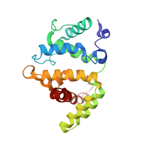
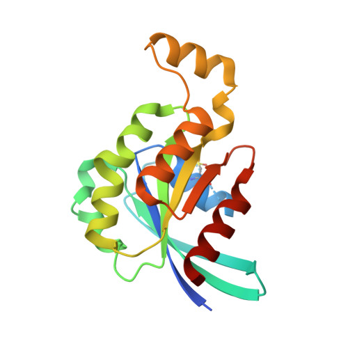
    RHO GTPase-activating proteins (RHOGAPs) are one of the major classes of regulators of the RHO-related protein family that are crucial in many cellular processes, motility, contractility, growth, differentiation, and development. Using database searches, we extracted 66 distinct human RHOGAPs, from which 57 have a common catalytic domain capable of terminating RHO protein signaling by stimulating the slow intrinsic GTP hydrolysis (GTPase) reaction. The specificity of the majority of the members of RHOGAP family is largely uncharacterized. Here, we comprehensively investigated the sequence-structure-function relationship between RHOGAPs and RHO proteins by combining our in vitro data with in silico data. The activity of 14 representatives of the RHOGAP family toward 12 RHO family proteins was determined in real time. We identified and structurally verified hot spots in the interface between RHOGAPs and RHO proteins as critical determinants for binding and catalysis. We have found that the RHOGAP domain itself is nonselective and in some cases rather inefficient under cell-free conditions. Thus, we propose that other domains of RHOGAPs confer substrate specificity and fine-tune their catalytic efficiency in cells.


  • Organizational Affiliation

    From the Institute of Biochemistry and Molecular Biology II, Medical Faculty, Heinrich-Heine-University, 40225 Düsseldorf, Germany.


Macromolecules
Find similar proteins by:  (by identity cutoff)  |  3D Structure
Entity ID: 1
MoleculeChains Sequence LengthOrganismDetailsImage
Rho GTPase-activating protein 35A,
C [auth B]
201Rattus norvegicusMutation(s): 0 
Gene Names: Arhgap35Grlf1
UniProt
Find proteins for P81128 (Rattus norvegicus)
Explore P81128 
Go to UniProtKB:  P81128
Entity Groups  
Sequence Clusters30% Identity50% Identity70% Identity90% Identity95% Identity100% Identity
UniProt GroupP81128
Sequence Annotations
Expand
  • Reference Sequence
Find similar proteins by:  (by identity cutoff)  |  3D Structure
Entity ID: 2
MoleculeChains Sequence LengthOrganismDetailsImage
Transforming protein RhoAB [auth F],
D
186Homo sapiensMutation(s): 1 
Gene Names: RHOAARH12ARHARHO12
UniProt & NIH Common Fund Data Resources
Find proteins for P61586 (Homo sapiens)
Explore P61586 
Go to UniProtKB:  P61586
PHAROS:  P61586
GTEx:  ENSG00000067560 
Entity Groups  
Sequence Clusters30% Identity50% Identity70% Identity90% Identity95% Identity100% Identity
UniProt GroupP61586
Sequence Annotations
Expand
  • Reference Sequence
Small Molecules
Ligands 4 Unique
IDChains Name / Formula / InChI Key2D Diagram3D Interactions
GDP
Query on GDP

Download Ideal Coordinates CCD File 
G [auth F],
K [auth D]
GUANOSINE-5'-DIPHOSPHATE
C10 H15 N5 O11 P2
QGWNDRXFNXRZMB-UUOKFMHZSA-N
MGF
Query on MGF

Download Ideal Coordinates CCD File 
I [auth F],
M [auth D]
TRIFLUOROMAGNESATE
F3 Mg
GJOMWUHGUQLOAC-UHFFFAOYSA-K
CL
Query on CL

Download Ideal Coordinates CCD File 
E [auth A],
F [auth A],
J [auth B]
CHLORIDE ION
Cl
VEXZGXHMUGYJMC-UHFFFAOYSA-M
MG
Query on MG

Download Ideal Coordinates CCD File 
H [auth F],
L [auth D]
MAGNESIUM ION
Mg
JLVVSXFLKOJNIY-UHFFFAOYSA-N
Modified Residues  1 Unique
IDChains TypeFormula2D DiagramParent
CSX
Query on CSX
A,
C [auth B]
L-PEPTIDE LINKINGC3 H7 N O3 SCYS
Experimental Data & Validation

Experimental Data

  • Method: X-RAY DIFFRACTION
  • Resolution: 1.72 Å
  • R-Value Free: 0.213 
  • R-Value Work: 0.170 
  • R-Value Observed: 0.171 
  • Space Group: P 21 21 21
Unit Cell:
Length ( Å )Angle ( ˚ )
a = 47.026α = 90
b = 112.322β = 90
c = 147.611γ = 90
Software Package:
Software NamePurpose
PHENIXrefinement
SERGUIdata collection
SCALEPACKdata scaling
PDB_EXTRACTdata extraction
BALBESphasing

Structure Validation

View Full Validation Report


Ligand Structure Quality Assessment 


Entry History & Funding Information

Deposition Data


Funding OrganizationLocationGrant Number
National Institutes of Health/National Institute of General Medical Sciences (NIH/NIGMS)United StatesR01GM086457

Revision History  (Full details and data files)

  • Version 1.0: 2016-08-17
    Type: Initial release
  • Version 1.1: 2016-10-05
    Changes: Database references
  • Version 1.2: 2017-09-20
    Changes: Author supporting evidence, Database references, Derived calculations
  • Version 1.3: 2019-12-25
    Changes: Author supporting evidence