5INH

Crystal structure of Autotaxin/ENPP2 with a covalent fragment


Experimental Data Snapshot

  • Method: X-RAY DIFFRACTION
  • Resolution: 1.84 Å
  • R-Value Free: 0.207 
  • R-Value Work: 0.172 
  • R-Value Observed: 0.174 

wwPDB Validation   3D Report Full Report


Ligand Structure Quality Assessment 


This is version 2.0 of the entry. See complete history


Literature

Repurposing Suzuki Coupling Reagents as a Directed Fragment Library Targeting Serine Hydrolases and Related Enzymes.

Lanier, M.Cole, D.C.Istratiy, Y.Klein, M.G.Schwartz, P.A.Tjhen, R.Jennings, A.Hixon, M.S.

(2017) J Med Chem 60: 5209-5215

  • DOI: https://doi.org/10.1021/acs.jmedchem.6b01224
  • Primary Citation of Related Structures:  
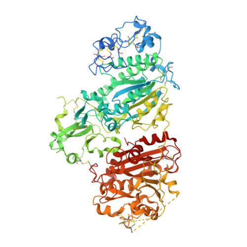
    5INH

  • PubMed Abstract: 

    Serine hydrolases are susceptible to potent reversible inhibition by boronic acids. Large collections of chemically diverse boronic acid fragments are commercially available because of their utility in coupling chemistry. We repurposed the approximately 650 boronic acid reagents in our collection as a directed fragment library targeting serine hydrolases and related enzymes. Highly efficient hits (LE > 0.6) often result. The utility of the approach is illustrated with the results against autotaxin, a phospholipase implicated in cardiovascular disease.


  • Organizational Affiliation

    Medicinal Chemistry - Gastrointestinal Drug Discovery Unit, ‡Structural Biology & Biophysics, §Modeling & Simulation-Global DMPK, Gastrointestinal Drug Discovery Unit, Takeda California, Inc. , 10410 Science Center Drive, San Diego, California 92121, United States.


Macromolecules
Find similar proteins by:  (by identity cutoff)  |  3D Structure
Entity ID: 1
MoleculeChains Sequence LengthOrganismDetailsImage
Ectonucleotide pyrophosphatase/phosphodiesterase family member 2879Mus musculusMutation(s): 0 
Gene Names: Enpp2Npps2Pdnp2
EC: 3.1.4.39
UniProt
Find proteins for Q9R1E6 (Mus musculus)
Explore Q9R1E6 
Go to UniProtKB:  Q9R1E6
Entity Groups  
Sequence Clusters30% Identity50% Identity70% Identity90% Identity95% Identity100% Identity
UniProt GroupQ9R1E6
Sequence Annotations
Expand
  • Reference Sequence
Oligosaccharides

Help

Entity ID: 2
MoleculeChains Length2D Diagram Glycosylation3D Interactions
alpha-D-mannopyranose-(1-2)-alpha-D-mannopyranose-(1-2)-alpha-D-mannopyranose-(1-3)-beta-D-mannopyranose-(1-4)-2-acetamido-2-deoxy-beta-D-glucopyranose-(1-4)-2-acetamido-2-deoxy-beta-D-glucopyranose
B
6N-Glycosylation
Glycosylation Resources
GlyTouCan:  G01760ZU
GlyCosmos:  G01760ZU
GlyGen:  G01760ZU
Entity ID: 3
MoleculeChains Length2D Diagram Glycosylation3D Interactions
2-acetamido-2-deoxy-beta-D-glucopyranose-(1-4)-2-acetamido-2-deoxy-beta-D-glucopyranose
C
2N-Glycosylation
Glycosylation Resources
GlyTouCan:  G42666HT
GlyCosmos:  G42666HT
GlyGen:  G42666HT
Small Molecules
Ligands 5 Unique
IDChains Name / Formula / InChI Key2D Diagram3D Interactions
6C1
Query on 6C1

Download Ideal Coordinates CCD File 
I [auth A]{2-[(3-chlorophenyl)methoxy]phenyl}boronic acid
C13 H12 B Cl O3
CYKRFBRLRUNSDG-UHFFFAOYSA-N
NAG
Query on NAG

Download Ideal Coordinates CCD File 
G [auth A]2-acetamido-2-deoxy-beta-D-glucopyranose
C8 H15 N O6
OVRNDRQMDRJTHS-FMDGEEDCSA-N
ZN
Query on ZN

Download Ideal Coordinates CCD File 
D [auth A],
E [auth A]
ZINC ION
Zn
PTFCDOFLOPIGGS-UHFFFAOYSA-N
ACT
Query on ACT

Download Ideal Coordinates CCD File 
H [auth A]ACETATE ION
C2 H3 O2
QTBSBXVTEAMEQO-UHFFFAOYSA-M
CA
Query on CA

Download Ideal Coordinates CCD File 
F [auth A]CALCIUM ION
Ca
BHPQYMZQTOCNFJ-UHFFFAOYSA-N
Binding Affinity Annotations 
IDSourceBinding Affinity
6C1 BindingDB:  5INH IC50: 1000 (nM) from 1 assay(s)
Experimental Data & Validation

Experimental Data

  • Method: X-RAY DIFFRACTION
  • Resolution: 1.84 Å
  • R-Value Free: 0.207 
  • R-Value Work: 0.172 
  • R-Value Observed: 0.174 
  • Space Group: P 1 21 1
Unit Cell:
Length ( Å )Angle ( ˚ )
a = 62.358α = 90
b = 80.745β = 102.86
c = 76.48γ = 90
Software Package:
Software NamePurpose
REFMACrefinement
HKL-2000data reduction
HKL-2000data scaling
PHASERphasing

Structure Validation

View Full Validation Report


Ligand Structure Quality Assessment 


Entry History 

Deposition Data

Revision History  (Full details and data files)

  • Version 1.0: 2017-03-15
    Type: Initial release
  • Version 1.1: 2017-06-21
    Changes: Database references
  • Version 1.2: 2017-07-05
    Changes: Database references
  • Version 2.0: 2020-07-29
    Type: Remediation
    Reason: Carbohydrate remediation
    Changes: Atomic model, Data collection, Derived calculations, Structure summary