5H2B

Structure of a novel antibody G196


Experimental Data Snapshot

  • Method: X-RAY DIFFRACTION
  • Resolution: 2.00 Å
  • R-Value Free: 0.281 
  • R-Value Work: 0.211 
  • R-Value Observed: 0.215 

wwPDB Validation   3D Report Full Report


This is version 1.2 of the entry. See complete history


Literature

G196 epitope tag system: a novel monoclonal antibody, G196, recognizes the small, soluble peptide DLVPR with high affinity.

Tatsumi, K.Sakashita, G.Nariai, Y.Okazaki, K.Kato, H.Obayashi, E.Yoshida, H.Sugiyama, K.Park, S.Y.Sekine, J.Urano, T.

(2017) Sci Rep 7: 43480-43480

  • DOI: https://doi.org/10.1038/srep43480
  • Primary Citation of Related Structures:  
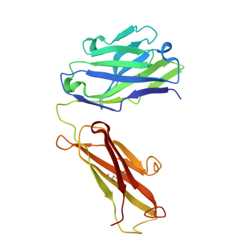
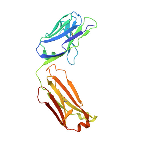
    5H2B

  • PubMed Abstract: 

    The recognition specificity of monoclonal antibodies (mAbs) has made mAbs among the most frequently used tools in both basic science research and in clinical diagnosis and therapies. Precise determination of the epitope allows the development of epitope tag systems to be used with recombinant proteins for various purposes. Here we describe a new family of tag derived from the epitope recognized by a highly specific mAb G196. The minimal epitope was identified as the five amino acid sequence Asp-Leu-Val-Pro-Arg. Permutation analysis was used to characterize the binding requirements of mAb G196, and the variable regions of the mAb G196 were identified and structurally analyzed by X-ray crystallography. Isothermal titration calorimetry revealed the high affinity (K d  = 1.25 nM) of the mAb G196/G196-epitope peptide interaction, and G196-tag was used to detect several recombinant cytosolic and nuclear proteins in human and yeast cells. mAb G196 is valuable for developing a new peptide tagging system for cell biology and biochemistry research.


  • Organizational Affiliation

    Department of Biochemistry, Shimane University School of Medicine, Izumo 693-8501, Japan.


Macromolecules
Find similar proteins by:  (by identity cutoff)  |  3D Structure
Entity ID: 1
MoleculeChains Sequence LengthOrganismDetailsImage
G196 antibody Heavy chain218Mus musculusMutation(s): 0 
Entity Groups  
Sequence Clusters30% Identity50% Identity70% Identity90% Identity95% Identity100% Identity
Sequence Annotations
Expand
  • Reference Sequence
Find similar proteins by:  (by identity cutoff)  |  3D Structure
Entity ID: 2
MoleculeChains Sequence LengthOrganismDetailsImage
G196 antibody Light chain214Mus musculusMutation(s): 0 
Entity Groups  
Sequence Clusters30% Identity50% Identity70% Identity90% Identity95% Identity100% Identity
Sequence Annotations
Expand
  • Reference Sequence
Experimental Data & Validation

Experimental Data

  • Method: X-RAY DIFFRACTION
  • Resolution: 2.00 Å
  • R-Value Free: 0.281 
  • R-Value Work: 0.211 
  • R-Value Observed: 0.215 
  • Space Group: P 21 21 21
Unit Cell:
Length ( Å )Angle ( ˚ )
a = 38.647α = 90
b = 82.267β = 90
c = 127.312γ = 90
Software Package:
Software NamePurpose
PHENIXrefinement
HKL-2000data reduction
SCALEPACKdata scaling
MOLREPphasing

Structure Validation

View Full Validation Report


Entry History 

Deposition Data

Revision History  (Full details and data files)

  • Version 1.0: 2017-03-22
    Type: Initial release
  • Version 1.1: 2017-04-19
    Changes: Structure summary
  • Version 1.2: 2020-02-26
    Changes: Data collection