5GOV

Crystal Structure of MCR-1, a phosphoethanolamine transferase, extracellular domain


Experimental Data Snapshot

  • Method: X-RAY DIFFRACTION
  • Resolution: 2.33 Å
  • R-Value Free: 0.236 
  • R-Value Work: 0.186 
  • R-Value Observed: 0.188 

wwPDB Validation   3D Report Full Report


This is version 1.2 of the entry. See complete history


Literature

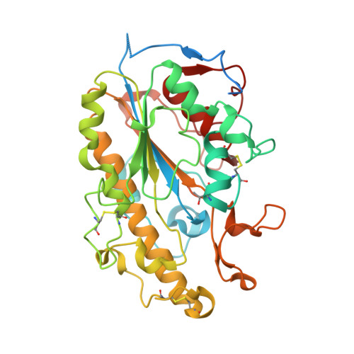
Crystal Structure of Escherichia coli originated MCR-1, a phosphoethanolamine transferase for Colistin Resistance.

Hu, M.Guo, J.Cheng, Q.Yang, Z.Chan, E.W.C.Chen, S.Hao, Q.

(2016) Sci Rep 6: 38793-38793

  • DOI: https://doi.org/10.1038/srep38793

  • PubMed Abstract: 

    MCR-1 is a phosphoethanolamine (pEtN) transferase that modifies the pEtN moiety of lipid A, conferring resistance to colistin, which is an antibiotic belonging to the class of polypeptide antibiotics known as polymyxins and is the last-line antibiotic used to treat multidrug resistant bacterial infections. Here we determined the crystal structure of the catalytic domain of MCR-1 (MCR-1-ED), which is originated in Escherichia coli (E. coli). MCR-1-ED was found to comprise several classical β-α-β-α motifs that constitute a "sandwich" conformation. Two interlaced molecules with different phosphorylation status of the residue T285 could give rise to two functional statuses of MCR-1 depending on the physiological conditions. MCR-1, like other known pEtN transferases, possesses an enzymatic site equipped with zinc binding residues. Interestingly, two zinc ions were found to mediate intermolecular interactions between MCR-1-ED molecules in one asymmetric unit and hence concatenation of MCR-1, allowing the protein to be oligomer. Findings of this work shall provide important insight into development of effective and clinically useful inhibitors of MCR-1 or structurally similar enzymes.


  • Organizational Affiliation

    School of Biomedical Sciences, University of Hong Kong, Laboratory Block, 21 Sassoon Road, Pokfulam, Hong Kong, China.


Macromolecules
Find similar proteins by:  (by identity cutoff)  |  3D Structure
Entity ID: 1
MoleculeChains Sequence LengthOrganismDetailsImage
Probable phosphatidylethanolamine transferase Mcr-1A [auth B],
B [auth A]
365Escherichia coliMutation(s): 0 
Gene Names: mcr1mcr-1
EC: 2.7
UniProt
Find proteins for A0A0R6L508 (Escherichia coli)
Explore A0A0R6L508 
Go to UniProtKB:  A0A0R6L508
Entity Groups  
Sequence Clusters30% Identity50% Identity70% Identity90% Identity95% Identity100% Identity
UniProt GroupA0A0R6L508
Sequence Annotations
Expand
  • Reference Sequence
Small Molecules
Ligands 1 Unique
IDChains Name / Formula / InChI Key2D Diagram3D Interactions
ZN
Query on ZN

Download Ideal Coordinates CCD File 
C [auth B]
D [auth B]
E [auth B]
F [auth B]
G [auth B]
C [auth B],
D [auth B],
E [auth B],
F [auth B],
G [auth B],
H [auth A],
I [auth A],
J [auth A],
K [auth A],
L [auth A],
M [auth A]
ZINC ION
Zn
PTFCDOFLOPIGGS-UHFFFAOYSA-N
Modified Residues  1 Unique
IDChains TypeFormula2D DiagramParent
TPO
Query on TPO
A [auth B],
B [auth A]
L-PEPTIDE LINKINGC4 H10 N O6 PTHR
Experimental Data & Validation

Experimental Data

  • Method: X-RAY DIFFRACTION
  • Resolution: 2.33 Å
  • R-Value Free: 0.236 
  • R-Value Work: 0.186 
  • R-Value Observed: 0.188 
  • Space Group: P 21 21 21
Unit Cell:
Length ( Å )Angle ( ˚ )
a = 54.439α = 90
b = 55.833β = 90
c = 219.388γ = 90
Software Package:
Software NamePurpose
PHENIXrefinement
HKL-2000data reduction
HKL-2000data scaling
PHASERphasing

Structure Validation

View Full Validation Report


Entry History 

Deposition Data

Revision History  (Full details and data files)

  • Version 1.0: 2016-12-21
    Type: Initial release
  • Version 1.1: 2020-01-01
    Changes: Advisory, Database references
  • Version 1.2: 2023-11-08
    Changes: Data collection, Database references, Derived calculations, Refinement description