5G5Y

S.pneumoniae ABC-transporter substrate binding protein FusA apo structure


Experimental Data Snapshot

  • Method: X-RAY DIFFRACTION
  • Resolution: 1.73 Å
  • R-Value Free: 0.193 
  • R-Value Work: 0.161 
  • R-Value Observed: 0.162 

wwPDB Validation   3D Report Full Report


This is version 1.0 of the entry. See complete history


Literature

Structural Basis for Regulation and Specificity of Fructooligosaccharide Import in Streptococcus pneumoniae.

Culurgioni, S.Harris, G.Singh, A.K.King, S.J.Walsh, M.A.

(2017) Structure 25: 79-93

  • DOI: https://doi.org/10.1016/j.str.2016.11.008
  • Primary Citation of Related Structures:  
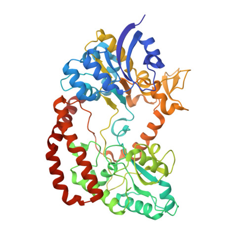
    5G5Y

  • PubMed Abstract: 

    Streptococcus pneumoniae is dependent on carbohydrate uptake for colonization and pathogenesis, and dedicates over a third of its transport systems to their uptake. The ability of the pneumococcus to utilize fructooligosaccharides (FOSs) is attributed to the presence of one of two types of FOS ATP-binding cassette (ABC) transporters. Strains encoding SfuABC are only able to utilize short-chain FOSs, while strains encoding FusABC can utilize both short- and long-chain FOSs. The crystal structures of the substrate-binding protein FusA in its open and closed conformations bound to FOSs, and solution scattering data of SfuA, delineate the structural basis for import of short- and long-chain FOSs. The structure of FusA identifies an EF hand-like calcium-binding motif. This is shown to be essential for translocation of FOSs in FusABC and forms the basis for the definition of a new class of substrate-binding proteins that regulate substrate translocation by calcium.


  • Organizational Affiliation

    Diamond House, Diamond Light Source, Harwell Science and Innovation Campus, Didcot OX11 0DE, UK; Research Complex at Harwell, Harwell Science and Innovation Campus, Didcot OX11 0FA, UK.


Macromolecules
Find similar proteins by:  (by identity cutoff)  |  3D Structure
Entity ID: 1
MoleculeChains Sequence LengthOrganismDetailsImage
ABC TRANSPORTER, SUBSTRATE-BINDING PROTEIN
A, B
494Streptococcus pneumoniae TIGR4Mutation(s): 0 
UniProt
Find proteins for A0A0H2URD6 (Streptococcus pneumoniae serotype 4 (strain ATCC BAA-334 / TIGR4))
Explore A0A0H2URD6 
Go to UniProtKB:  A0A0H2URD6
Entity Groups  
Sequence Clusters30% Identity50% Identity70% Identity90% Identity95% Identity100% Identity
UniProt GroupA0A0H2URD6
Sequence Annotations
Expand
  • Reference Sequence
Small Molecules
Ligands 4 Unique
IDChains Name / Formula / InChI Key2D Diagram3D Interactions
SO4
Query on SO4

Download Ideal Coordinates CCD File 
E [auth A]SULFATE ION
O4 S
QAOWNCQODCNURD-UHFFFAOYSA-L
GOL
Query on GOL

Download Ideal Coordinates CCD File 
M [auth A],
N [auth A],
O [auth A],
U [auth B],
V [auth B]
GLYCEROL
C3 H8 O3
PEDCQBHIVMGVHV-UHFFFAOYSA-N
ZN
Query on ZN

Download Ideal Coordinates CCD File 
F [auth A]
G [auth A]
H [auth A]
I [auth A]
J [auth A]
F [auth A],
G [auth A],
H [auth A],
I [auth A],
J [auth A],
K [auth A],
L [auth A],
R [auth B],
S [auth B],
T [auth B]
ZINC ION
Zn
PTFCDOFLOPIGGS-UHFFFAOYSA-N
CA
Query on CA

Download Ideal Coordinates CCD File 
C [auth A],
D [auth A],
P [auth B],
Q [auth B]
CALCIUM ION
Ca
BHPQYMZQTOCNFJ-UHFFFAOYSA-N
Experimental Data & Validation

Experimental Data

  • Method: X-RAY DIFFRACTION
  • Resolution: 1.73 Å
  • R-Value Free: 0.193 
  • R-Value Work: 0.161 
  • R-Value Observed: 0.162 
  • Space Group: P 1
Unit Cell:
Length ( Å )Angle ( ˚ )
a = 66.07α = 108.02
b = 72.17β = 104.99
c = 77.66γ = 92.49
Software Package:
Software NamePurpose
REFMACrefinement
XDSdata reduction
SCALAdata scaling
PHASERphasing

Structure Validation

View Full Validation Report


Entry History 

Deposition Data

Revision History  (Full details and data files)

  • Version 1.0: 2017-01-18
    Type: Initial release