5FP0

ligand complex structure of soluble epoxide hydrolase


Experimental Data Snapshot

  • Method: X-RAY DIFFRACTION
  • Resolution: 2.35 Å
  • R-Value Free: 0.230 
  • R-Value Work: 0.170 
  • R-Value Observed: 0.173 

wwPDB Validation   3D Report Full Report


Ligand Structure Quality Assessment 


This is version 1.1 of the entry. See complete history


Literature

Fragment Screening of Soluble Epoxide Hydrolase for Lead Generation-Structure-Based Hit Evaluation and Chemistry Exploration.

Xue, Y.Olsson, T.Johansson, C.A.Oster, L.Beisel, H.Rohman, M.Karis, D.Backstrom, S.

(2016) ChemMedChem 11: 497

  • DOI: https://doi.org/10.1002/cmdc.201500575
  • Primary Citation of Related Structures:  
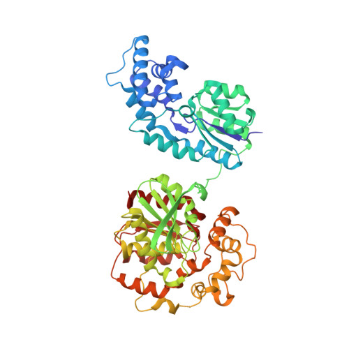
    5FP0

  • PubMed Abstract: 

    Soluble epoxide hydrolase (sEH) is involved in the regulation of many biological processes by metabolizing the key bioactive lipid mediator, epoxyeicosatrienoic acids. For the development of sEH inhibitors with improved physicochemical properties, we performed both a fragment screening and a high-throughput screening aiming at an integrated hit evaluation and lead generation. Followed by a joint dose-response analysis to confirm the hits, the identified actives were then effectively triaged by a structure-based hit-classification approach to three prioritized series. Two distinct scaffolds were identified as tractable starting points for potential lead chemistry work. The oxoindoline series bind at the right-hand side of the active-site pocket with hydrogen bonds to the protein. The 2-phenylbenzimidazole-4-sulfonamide series bind at the central channel with significant induced fit, which has not been previously reported. On the basis of the encouraging initial results, we envision that a new lead series with improved properties could be generated if a vector is found that could merge the cyclohexyl functionality of the oxoindoline series with the trifluoromethyl moiety of the 2-phenylbenzimidazole-4-sulfonamide series.


  • Organizational Affiliation

    Department Discovery Sciences, AstraZeneca R&D Gothenburg, Pepparedsleden 1, 431 83, Mölndal, Sweden.


Macromolecules
Find similar proteins by:  (by identity cutoff)  |  3D Structure
Entity ID: 1
MoleculeChains Sequence LengthOrganismDetailsImage
BIFUNCTIONAL EPOXIDE HYDROLASE 2549Homo sapiensMutation(s): 0 
EC: 3.3.2.10 (PDB Primary Data), 3.1.3.76 (PDB Primary Data)
UniProt & NIH Common Fund Data Resources
Find proteins for P34913 (Homo sapiens)
Explore P34913 
Go to UniProtKB:  P34913
PHAROS:  P34913
GTEx:  ENSG00000120915 
Entity Groups  
Sequence Clusters30% Identity50% Identity70% Identity90% Identity95% Identity100% Identity
UniProt GroupP34913
Sequence Annotations
Expand
  • Reference Sequence
Small Molecules
Ligands 2 Unique
IDChains Name / Formula / InChI Key2D Diagram3D Interactions
SZC
Query on SZC

Download Ideal Coordinates CCD File 
C [auth A]N-cyclopentyl-2-[4-(trifluoromethyl)phenyl]-3H-benzimidazole-4-sulfonamide
C19 H18 F3 N3 O2 S
BCAADIRANQOVRJ-UHFFFAOYSA-N
DMS
Query on DMS

Download Ideal Coordinates CCD File 
B [auth A]DIMETHYL SULFOXIDE
C2 H6 O S
IAZDPXIOMUYVGZ-UHFFFAOYSA-N
Binding Affinity Annotations 
IDSourceBinding Affinity
SZC Binding MOAD:  5FP0 IC50: 17 (nM) from 1 assay(s)
Experimental Data & Validation

Experimental Data

  • Method: X-RAY DIFFRACTION
  • Resolution: 2.35 Å
  • R-Value Free: 0.230 
  • R-Value Work: 0.170 
  • R-Value Observed: 0.173 
  • Space Group: P 65 2 2
Unit Cell:
Length ( Å )Angle ( ˚ )
a = 92.51α = 90
b = 92.51β = 90
c = 244.83γ = 120
Software Package:
Software NamePurpose
BUSTERrefinement
XDSdata reduction
SCALAdata scaling

Structure Validation

View Full Validation Report


Ligand Structure Quality Assessment 


Entry History 

Deposition Data

Revision History  (Full details and data files)

  • Version 1.0: 2016-03-02
    Type: Initial release
  • Version 1.1: 2016-03-16
    Changes: Database references