Crystal structure of Rv2258c from Mycobacterium tuberculosis H37Rv, an S-adenosyl-l-methionine-dependent methyltransferase
Im, H.N., Kim, H.S., An, D.R., Jang, J.Y., Kim, J., Yoon, H.J., Yang, J.K., Suh, S.W.(2016) J Struct Biol 193: 172-180
- PubMed: 26772148
- DOI: https://doi.org/10.1016/j.jsb.2016.01.002
- Primary Citation of Related Structures:
5F8C, 5F8E, 5F8F - PubMed Abstract:
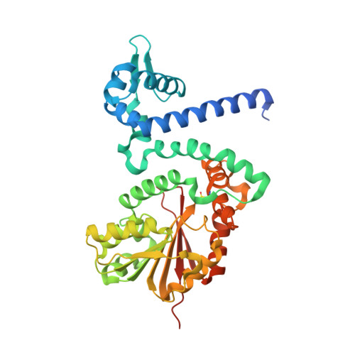
The Mycobacterium tuberculosis Rv2258c protein is an S-adenosyl-L-methionine (SAM)-dependent methyltransferase (MTase). Here, we have determined its crystal structure in three forms: a ligand-unbound form, a binary complex with sinefungin (SFG), and a binary complex with S-adenosyl-L-homocysteine (SAH). The monomer structure of Rv2258c consists of two domains which are linked by a long α-helix. The N-terminal domain is essential for dimerization and the C-terminal domain has the Class I MTase fold. Rv2258c forms a homodimer in the crystal, with the N-terminal domains facing each other. It also exists as a homodimer in solution. A DALI structural similarity search with Rv2258c reveals that the overall structure of Rv2258c is very similar to small-molecule SAM-dependent MTases. Rv2258c interacts with the bound SFG (or SAH) in an extended conformation maintained by a network of hydrogen bonds and stacking interactions. Rv2258c has a relatively large hydrophobic cavity for binding of the methyl-accepting substrate, suggesting that bulky nonpolar molecules with aromatic rings might be targeted for methylation by Rv2258c in M. tuberculosis. However, the ligand-binding specificity and the biological role of Rv2258c remain to be elucidated due to high variability of the amino acid residues defining the substrate-binding site.
Organizational Affiliation:
Department of Biophysics and Chemical Biology, College of Natural Sciences, Seoul National University, Seoul 08826, Republic of Korea.


