5EQ3

Crystal structure of the SrpA adhesin from Streptococcus sanguinis with a sialyl galactose disaccharide bound

  • Classification: SUGAR BINDING PROTEIN
  • Organism(s): Streptococcus sanguinis SK36
  • Expression System: Escherichia coli
  • Mutation(s): No 

  • Deposited: 2015-11-12 Released: 2016-01-27 
  • Deposition Author(s): Loukachevitch, L.V., McCulloch, K.M., Vann, K.R., Wawrzak, Z., Anderson, S., Iverson, T.M.
  • Funding Organization(s): National Institutes of Health/National Institute Of Allergy and Infectious Diseases (NIH/NIAID), Department of Veteran's Affairs, American Heart Association, National Institutes of Health/National Institute of General Medical Sciences (NIH/NIGMS), National Institutes of Health/National Heart, Lung, and Blood Institute (NIH/NHLBI), National Institutes of Health/National Center for Research Resources (NIH/NCRR), Department of Energy (DOE, United States), Michigan Economic Development Corporation

Experimental Data Snapshot

  • Method: X-RAY DIFFRACTION
  • Resolution: 2.00 Å
  • R-Value Free: 0.243 
  • R-Value Work: 0.198 
  • R-Value Observed: 0.200 

wwPDB Validation   3D Report Full Report


This is version 2.1 of the entry. See complete history


Literature

Structural Basis for Sialoglycan Binding by the Streptococcus sanguinis SrpA Adhesin.

Bensing, B.A.Loukachevitch, L.V.McCulloch, K.M.Yu, H.Vann, K.R.Wawrzak, Z.Anderson, S.Chen, X.Sullam, P.M.Iverson, T.M.

(2016) J Biol Chem 291: 7230-7240

  • DOI: https://doi.org/10.1074/jbc.M115.701425
  • Primary Citation of Related Structures:  
    5EQ2, 5EQ3, 5EQ4

  • PubMed Abstract: 

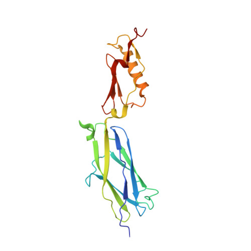
    Streptococcus sanguinisis a leading cause of infective endocarditis, a life-threatening infection of the cardiovascular system. An important interaction in the pathogenesis of infective endocarditis is attachment of the organisms to host platelets.S. sanguinisexpresses a serine-rich repeat adhesin, SrpA, similar in sequence to platelet-binding adhesins associated with increased virulence in this disease. In this study, we determined the first crystal structure of the putative binding region of SrpA (SrpABR) both unliganded and in complex with a synthetic disaccharide ligand at 1.8 and 2.0 Å resolution, respectively. We identified a conserved Thr-Arg motif that orients the sialic acid moiety and is required for binding to platelet monolayers. Furthermore, we propose that sequence insertions in closely related family members contribute to the modulation of structural and functional properties, including the quaternary structure, the tertiary structure, and the ligand-binding site.


  • Organizational Affiliation

    From the Division of Infectious Diseases, Veterans Affairs Medical Center, Department of Medicine, University of California, San Francisco and the Northern California Institute for Research and Education, San Francisco, California 94121.


Macromolecules
Find similar proteins by:  (by identity cutoff)  |  3D Structure
Entity ID: 1
MoleculeChains Sequence LengthOrganismDetailsImage
Platelet-binding glycoprotein
A, B
201Streptococcus sanguinis SK36Mutation(s): 0 
Gene Names: srpASSA_0829
UniProt
Find proteins for A3CM52 (Streptococcus sanguinis (strain SK36))
Explore A3CM52 
Go to UniProtKB:  A3CM52
Entity Groups  
Sequence Clusters30% Identity50% Identity70% Identity90% Identity95% Identity100% Identity
UniProt GroupA3CM52
Sequence Annotations
Expand
  • Reference Sequence
Oligosaccharides

Help

Entity ID: 2
MoleculeChains Length2D Diagram Glycosylation3D Interactions
N-glycolyl-alpha-neuraminic acid-(2-3)-methyl beta-D-galactopyranoside
C
2N/A
Glycosylation Resources
GlyTouCan:  G90552PC
GlyCosmos:  G90552PC
Small Molecules
Ligands 3 Unique
IDChains Name / Formula / InChI Key2D Diagram3D Interactions
ACT
Query on ACT

Download Ideal Coordinates CCD File 
G [auth A],
H [auth A],
N [auth B]
ACETATE ION
C2 H3 O2
QTBSBXVTEAMEQO-UHFFFAOYSA-M
CA
Query on CA

Download Ideal Coordinates CCD File 
D [auth A]
E [auth A]
F [auth A]
I [auth B]
J [auth B]
D [auth A],
E [auth A],
F [auth A],
I [auth B],
J [auth B],
K [auth B],
L [auth B]
CALCIUM ION
Ca
BHPQYMZQTOCNFJ-UHFFFAOYSA-N
NA
Query on NA

Download Ideal Coordinates CCD File 
M [auth B]SODIUM ION
Na
FKNQFGJONOIPTF-UHFFFAOYSA-N
Experimental Data & Validation

Experimental Data

Unit Cell:
Length ( Å )Angle ( ˚ )
a = 174.62α = 90
b = 47.005β = 102.15
c = 64.026γ = 90
Software Package:
Software NamePurpose
HKL-2000data collection
HKL-2000data scaling
PHENIXrefinement
PDB_EXTRACTdata extraction
HKL-2000data reduction

Structure Validation

View Full Validation Report


Entry History & Funding Information

Deposition Data


Funding OrganizationLocationGrant Number
National Institutes of Health/National Institute Of Allergy and Infectious Diseases (NIH/NIAID)United StatesAI106987
National Institutes of Health/National Institute Of Allergy and Infectious Diseases (NIH/NIAID)United StatesAI41513
Department of Veteran's AffairsUnited States--
American Heart AssociationUnited States14GRNT20390021
National Institutes of Health/National Institute of General Medical Sciences (NIH/NIGMS)United StatesT32 GM008320
National Institutes of Health/National Heart, Lung, and Blood Institute (NIH/NHLBI)United StatesT32 HL007751
National Institutes of Health/National Center for Research Resources (NIH/NCRR)United StatesS10 RR026915
Department of Energy (DOE, United States)United StatesDE-AC02-06CH11357
Michigan Economic Development CorporationUnited States085P1000817

Revision History  (Full details and data files)

  • Version 1.0: 2016-01-27
    Type: Initial release
  • Version 1.1: 2016-02-17
    Changes: Database references
  • Version 1.2: 2017-09-20
    Changes: Author supporting evidence, Derived calculations
  • Version 1.3: 2019-12-04
    Changes: Author supporting evidence
  • Version 2.0: 2020-07-29
    Type: Remediation
    Reason: Carbohydrate remediation
    Changes: Atomic model, Data collection, Derived calculations, Structure summary
  • Version 2.1: 2024-03-06
    Changes: Data collection, Database references, Derived calculations, Structure summary