


Structure of 3-dehydroquinate synthase from Acinetobacter baumannii in complex with NAD
Abendroth, J., Dranow, D.M., Lorimer, D.D., Edwards, T.E.To be published.
Experimental Data Snapshot
Entity ID: 1 | |||||
|---|---|---|---|---|---|
| Molecule | Chains | Sequence Length | Organism | Details | Image |
| 3-dehydroquinate synthase | 368 | Acinetobacter baumannii | Mutation(s): 0 Gene Names: aroB, AB895_1177, AB988_0958, AB994_2838, ABCIP7010_3378, ABUW_0296, ACX61_01585, IOMTU433_0312, RU84_01465, TE32_01480... EC: 4.2.3.4 | 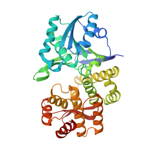 | |
UniProt | |||||
Find proteins for V5V8R5 (Acinetobacter baumannii) Explore V5V8R5 Go to UniProtKB: V5V8R5 | |||||
Entity Groups | |||||
| Sequence Clusters | 30% Identity50% Identity70% Identity90% Identity95% Identity100% Identity | ||||
| UniProt Group | V5V8R5 | ||||
Sequence AnnotationsExpand | |||||
| |||||
| Ligands 2 Unique | |||||
|---|---|---|---|---|---|
| ID | Chains | Name / Formula / InChI Key | 2D Diagram | 3D Interactions | |
| NAD Query on NAD | C [auth A], E [auth B] | NICOTINAMIDE-ADENINE-DINUCLEOTIDE C21 H27 N7 O14 P2 BAWFJGJZGIEFAR-NNYOXOHSSA-N |  | ||
| MG Query on MG | D [auth A], F [auth B] | MAGNESIUM ION Mg JLVVSXFLKOJNIY-UHFFFAOYSA-N |  | ||
| Length ( Å ) | Angle ( ˚ ) |
|---|---|
| a = 58.4 | α = 90 |
| b = 58.92 | β = 97.4 |
| c = 104.81 | γ = 90 |
| Software Name | Purpose |
|---|---|
| XDS | data reduction |
| XSCALE | data scaling |
| PHASER | phasing |
| ARP | model building |
| Coot | model building |
| PHENIX | refinement |
| PDB_EXTRACT | data extraction |
RCSB PDB (citation) is hosted by
RCSB PDB is a member of the

