5EJC

Crystal structural of the TSC1-TBC1D7 complex


Experimental Data Snapshot

  • Method: X-RAY DIFFRACTION
  • Resolution: 3.10 Å
  • R-Value Free: 0.239 
  • R-Value Work: 0.220 
  • R-Value Observed: 0.221 

wwPDB Validation   3D Report Full Report


This is version 1.4 of the entry. See complete history


Literature

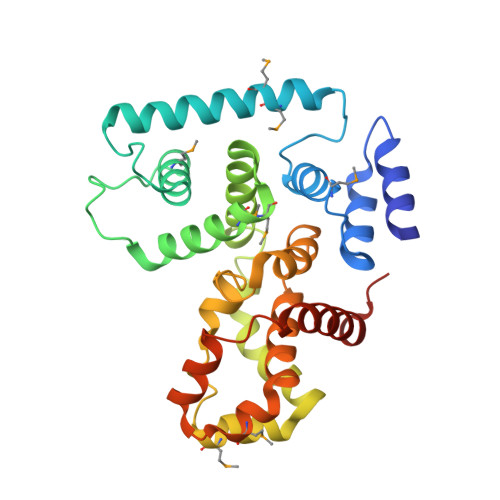
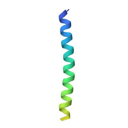
Structural Basis of the Interaction between Tuberous Sclerosis Complex 1 (TSC1) and Tre2-Bub2-Cdc16 Domain Family Member 7 (TBC1D7).

Qin, J.Wang, Z.Hoogeveen-Westerveld, M.Shen, G.Gong, W.Nellist, M.Xu, W.

(2016) J Biol Chem 291: 8591-8601

  • DOI: https://doi.org/10.1074/jbc.M115.701870
  • Primary Citation of Related Structures:  
    5EJC

  • PubMed Abstract: 

    Mutations in TSC1 or TSC2 cause tuberous sclerosis complex (TSC), an autosomal dominant disorder characterized by the occurrence of benign tumors in various vital organs and tissues. TSC1 and TSC2, the TSC1 and TSC2 gene products, form the TSC protein complex that senses specific cellular growth conditions to control mTORC1 signaling. TBC1D7 is the third subunit of the TSC complex, and helps to stabilize the TSC1-TSC2 complex through its direct interaction with TSC1. Homozygous inactivation of TBC1D7 causes intellectual disability and megaencephaly. Here we report the crystal structure of a TSC1-TBC1D7 complex and biochemical characterization of the TSC1-TBC1D7 interaction. TBC1D7 interacts with the C-terminal region of the predicted coiled-coil domain of TSC1. The TSC1-TBC1D7 interface is largely hydrophobic, involving the α4 helix of TBC1D7. Each TBC1D7 molecule interacts simultaneously with two parallel TSC1 helices from two TSC1 molecules, suggesting that TBC1D7 may stabilize the TSC complex by tethering the C-terminal ends of two TSC1 coiled-coils.


  • Organizational Affiliation

    From the Institute of Biophysics, Chinese Academy of Sciences, Beijing 100101, China, the Department of Biological Structure, University of Washington, Seattle, Washington 98195, the University of Chinese Academy of Sciences, Beijing 100049, China.


Macromolecules
Find similar proteins by:  (by identity cutoff)  |  3D Structure
Entity ID: 1
MoleculeChains Sequence LengthOrganismDetailsImage
TBC1 domain family member 7
A, B
276Homo sapiensMutation(s): 0 
Gene Names: TBC1D7TBC7HSPC239
UniProt & NIH Common Fund Data Resources
Find proteins for Q9P0N9 (Homo sapiens)
Explore Q9P0N9 
Go to UniProtKB:  Q9P0N9
PHAROS:  Q9P0N9
GTEx:  ENSG00000145979 
Entity Groups  
Sequence Clusters30% Identity50% Identity70% Identity90% Identity95% Identity100% Identity
UniProt GroupQ9P0N9
Sequence Annotations
Expand
  • Reference Sequence
Find similar proteins by:  (by identity cutoff)  |  3D Structure
Entity ID: 2
MoleculeChains Sequence LengthOrganismDetailsImage
Hamartin
C, D, E, F
55Homo sapiensMutation(s): 0 
Gene Names: TSC1KIAA0243TSC
UniProt & NIH Common Fund Data Resources
Find proteins for Q92574 (Homo sapiens)
Explore Q92574 
Go to UniProtKB:  Q92574
PHAROS:  Q92574
GTEx:  ENSG00000165699 
Entity Groups  
Sequence Clusters30% Identity50% Identity70% Identity90% Identity95% Identity100% Identity
UniProt GroupQ92574
Sequence Annotations
Expand
  • Reference Sequence
Small Molecules
Modified Residues  1 Unique
IDChains TypeFormula2D DiagramParent
MSE
Query on MSE
A, B
L-PEPTIDE LINKINGC5 H11 N O2 SeMET
Experimental Data & Validation

Experimental Data

  • Method: X-RAY DIFFRACTION
  • Resolution: 3.10 Å
  • R-Value Free: 0.239 
  • R-Value Work: 0.220 
  • R-Value Observed: 0.221 
  • Space Group: C 1 2 1
Unit Cell:
Length ( Å )Angle ( ˚ )
a = 183.395α = 90
b = 66.704β = 91.91
c = 98.37γ = 90
Software Package:
Software NamePurpose
REFMACrefinement
HKL-2000data scaling
PDB_EXTRACTdata extraction
HKL-2000data reduction
AutoSolphasing

Structure Validation

View Full Validation Report


Entry History & Funding Information

Deposition Data


Funding OrganizationLocationGrant Number
Department of Defense (DOD, United States)United StatesW81XWH-13-1-0059 (TS120073)

Revision History  (Full details and data files)

  • Version 1.0: 2016-03-02
    Type: Initial release
  • Version 1.1: 2016-03-09
    Changes: Database references
  • Version 1.2: 2016-05-11
    Changes: Database references
  • Version 1.3: 2019-04-10
    Changes: Author supporting evidence, Data collection, Database references, Derived calculations
  • Version 1.4: 2019-11-27
    Changes: Author supporting evidence